The bird leg anatomy exhibits several specialized features compared to that of mammals. They are using for weight-bearing, scratching, climbing, grasping, and swimming. In this article, I will show you the peculiar anatomical facts of bird legs with labeled diagrams. You will get detailed information on bird leg anatomy bones and bird leg muscle anatomy.
I will also describe the anatomy of the blood vessels (artery and vein) and nerves from the bird’s leg. In addition, you will find the answers to the frequently asked questions on birds legs at the end of this article.
Bird leg anatomy
You know the term leg means the part of any animal’s lower extremity that runs from the stifle joint to the hock joint. So, the bird leg anatomy will include only the tibia, fibula bones, and associated structures. But, here, I will describe all the bones from the pelvic limb of a bird.
I will also describe the anatomical facts of all clinically important muscles from the birds legs. The most important vein of the bird leg is the shank vein (caudal tibial). If you want to collect blood from a bird for diagnosis purposes, you might know the anatomy of this vein.
“It is not good practice to collect blood from the bird caudal tibial vein. You may collect blood from the bird wing vein (subclavian vein), which is easier than the shank vein. Please don’t practice this method if you are not a registered veterinarian.”
The sciatic nerve of the bird leg is the clinically important and largest nerve in the body. You should know the courses and normal appearance of the sciatic nerve of the bird leg. In some diseases, you may find some unique lessons on the bird sciatic nerve.
So, from the birds legs, You might learn the below-mentioned structures –
- Anatomical facts of all bones from the birds leg (or pelvic limb)
- The muscle anatomy of the bird leg,
- A short course of the clinically important blood vessels, and
- The clinically important larger sciatic nerve of a bird
You will find a great variation in the digits and claws of a bird compared to mammals. I hope you will find it interest to learn these interesting facts about the birds legs.
Bird leg anatomy bones
So, the bones of a bird leg anatomy include the femur, tibiotarsus, fibula, tarsometatarsus, and phalanges. Before going to the detailed description of these legs bones, I would like to provide a little information about the pelvic girdle.
The pelvic girdle of a bird consists of the three bones – ilium, ischium, and pubis. They are anatomically different from mammals.
These bones have a rigid connection and form the ox coxae in the bird skeleton. They also have a connection with the synsacrum of a bird skeleton.
If you want to know the summary of the osteological features of all the bones from a bird skeleton, you may read the following article –
The bird or chicken skeleton anatomy with a labeled diagram
You will also find the videos and labeled diagrams related to the bird skeleton on the social media of anatomylearner.com.
There is no pubic symphysis in the bird pelvic girdle. So, the bird’s pelvis is open ventrally, and it facilitates the passages of the egg.
Bones of bird pelvic girdle
The osteological features of the bird pelvic girdle bones are almost similar to those of a chicken. There is no actual boundary of these three bones in the bird pelvic girdle. However, you may identify these bones with the help of the following information.
Ilium of the bird pelvic girdle
This is the largest bone of the pelvic girdle that forms the dorsal surface of the bony pelvis. The bird ilium bone has three main divisions – the preacetabular wing, a body, and a postacetabular wing.
The preacetabular wing of the bird surrounds by the dorsal iliac crest dorsally. Again, this crest connects with the spinous processes of the synsacrum and form the iliosynsacral crest.
You will also find the iliosynsacral canal on the dorsal surface of the ilium bone of a bird. In addition, there is a lateral iliac crest that surrounds the craniolateral margin of the preacetabular wing of a bird. Here, you will find an expanded area for the attachment of the muscle of the bird’s legs.
The lateral surface of the ilium bone joins with the wing of the ischium bone without any distinct boundary. At the ventral surface of the bird ilium, there is a bony crypt for the caudal division of the kidney.
The body of the bird ilium locates craniodorsally of the bone. You will find the preacetabular tubercle and antitrochanter that arises from the bird ilium body.
In the postacetabular wing of a bird ilium, you will find two portions – dorsal and lateral surfaces. The dorsal surface has a connection with the synsacrum and separates from the lateral surface by the dorsolateral iliac crest.
Again, the dorsolateral iliac crest continues caudally of the postacetabular wing. Then it tappers and form the dorsolateral iliac spine of the postacetabular wing.
Ischium and pubis of bird pelvic girdle
In the bird ischium bone, you will find a body and a wing. The body of the bird ischium bone contributes to forming the acetabulum cavity caudally. It also contributes to forming the part of the ventral antitrochanter.
The wing of the bird ischium bone locates at the lateral position. It forms the ventral continuation of the lateral surface of the ilium bone. You will not find any distinctive osteological features in the bird ischium bone. But, it serves as the origin of the muscles of the pelvic limb of a bird.
The pubis of a bird is the most ventral bone of the pelvic girdle. It contributes to forming the ventral boundary of the acetabulum cavity. Then it continues as a rod-shaped structure caudally and remains incomplete fusion with the ischium bone.
This pubis bone also contributes to forming the obturator foramen cranially and ischiopubic window caudally.
A femur from bird leg anatomy
The femur is the first long bone in the bird leg anatomy. It is cylindrical and has a slight cranial curvature. You will find two extremities and a body in the bird femur. Now, I will show you all the anatomical facts of the bird femur with a labeled diagram.
The body of the bird femur is cylindrical and smooth side to side. But, you may find some muscular lines on the cranial and caudal surface of the femur.
Extremities of the bird femur bone
The proximal extremity of the bird femur possesses a rounded femoral head. This femoral head also posses a small cavity for the ligament of the femoral head.
There is a small constricted neck that separates the head from the body of the femur bone. You might find an articular surface for the joint with an antitraochenter structure.
Again, at the proximal extremity, you will also find a distinct trochanteric femoris. There is a large trochanteric crest run along with the trochanteric femoris. This trochanteric crest runs cranially and ventrally of the bird femur.
In addition, the distal extremity of the bird femur bone posses the lateral and medial condyle. You will also find the medial and lateral epicondyle just above these condyles. The intercondylar groove separates the lateral and medial condyles of the bird femur.
There is a circular fibular notch at the lateral surface of the lateral condyle of the bird femur. This fibular notch articulates with the fibula of a bird.
Again, at the distal extremity of the bird femur, you will find a patellar groove in the cranial surface. It bounds by the bony crest on both sides. In addition, this groove provides the gliding articular surface for the patella.
Patella of the bird legs
The patella is also the largest sesamoid bone in bird legs as in mammals. It lines with the cartilage on its caudally directed articular surface. You will find a groove on the cranial surface of the bird patella.
The patella of a bird is not so well-developed as in mammals. It is a thin triangular bone-like structure in a bird. The width is comparatively more than the length.
Tibiotarsus of the bird leg anatomy
The leg bones of the proximal row of the tarsus are fused with the distal end of the bird tibia. Hence, another name of this bone is tibiotarsus. In the bird leg anatomy, the tibiotarsus is the longest bone that posses a prominent tibial crest.
Here, I will show you the osteological features from the proximal extremity, body, and distal extremity. At the proximal extremity of the tibiotarsus bone, you will find the lateral and medial articular surface that separates by the interarticular area.
The most prominent feature of the bird tibiotarsus is the large tibial crest (cnemial crest). You will also find the patellar and lateral crest in the proximal extremity of the bird tibiotarsus bone. The patellar crest joins with the lateral crest of the tibiotarsus.
In addition, you will also find a large intercnemial groove at the lateral aspect of the proximal tibiotarsus.
At the distal extremity, there are lateral and medial condyles present in the tibiotarsus. As well as, you will also find the lateral and medial epicondyles in the distal extremity of the bird tibiotarsus bone.
An articular surface (trochlea for tibial cartilage) is present at the caudal aspect of the distal tibiotarsus bone. At the cranial aspect of the distal tibiotarsus, you will find the below-mentioned unique structures –
An extensor groove of the tibiotarsus
The supratendinal bridge of the tibiotarsus
An intercondyloid fossa of the distal tibiotarsus
The extensor groove locates just proximal to the distal extremity of the bird tibiotarsus bone.
Bird fibula bone anatomy
The fibula is the thin rod-shaped long bone attaches to the lateral part of the proximal end of the tibiotarsus bone. You will find a head and a slender body in the bird fibula bone.
The head of the bird fibula posses two articular surfaces – one for the tibiotarsus and another for the lateral condyle of the femur.
Tarsometatarsus from the bird leg anatomy
In the tarsometatarsus of the bird leg anatomy, you will find unique osteological features than mammals. The tarsometatarsus bone forms by fusing the central and distal tarsal with the metatarsal II, III, and IV. You will also find a small metatarsal bone in the bird legs located at the distal end’s posterior aspect.
At the proximal end of the tarsometatarsus bone, two articular surfaces are separated by the intercondylar eminence. Within these articular surfaces, the tibiotarsus articulate.
Again, there is a hypotarsus at the caudal aspect of the proximal end of a bird tarsometatarsal. It contains a longitudinally oriented crest and channels.
At the distal end, the tarsometatarsal bone posses three trochleae for articulation with the digits II, III, and IV. The metatarsal bone I also possess a small trochlea on itsdistomedial aspect.
You will find a boy structure that arises from the medial aspect of the body of the tarsometatarsus bone. This is the spur, and it commonly occurs in the male bird legs.
Phalanges and digits of the bird leg
In the bird digits, you will find different numbers of phalanges. The first digit of the bird posses only two phalanges. Again, the number of the phalanges increases by one in the next digits. So, you will find three phalanges in the second digit of a bird. In addition, the third digit possesses four, and the fourth digit posses five phalanges.
There are three types of phalanges present in the bird digits – proximal, intermediate, and distal. The osteological features of the proximal and intermediate phalanges are almost similar. You will find a base with an articular surface, a body, and a capitulum in both proximal and intermediate phalanges.
But the distal phalanges possess a base and an apex. You will also find a bony structure (claw) at the tip of the distal phalange of a birds leg.
Joints of the bird leg anatomy
Here, I will not describe the detailed anatomy of the bird leg joints. I will only provide a piece of basic information on all the joints of the birds legs. Fine, if you want to learn the details anatomy of the bird hindlimb joint, you may read the other article from anatomylearner.com.
The os coxae of the bird have a rigid connection with the synsacrum structure. You will also find a rigid connection among the three bones of the bird os coxae (except in pubis).
In the bird hind leg, you will find the following joints –
- The hip joint of the bird leg
- A knee joint of the bird leg
- The intertarsal joint of the bird leg,
- Joints of the bird metatarsal bones
- The joints of the metatarsal and phalanges
Let’s know some of the important joint structures (bones and ligaments involvement) from the birds leg.
The hip joint of the bird hind leg
In the bird hip joint, the head of femur articulate with the acetabular cavity. This joint comprises two articulations – the coxocapital joint and the coxotrochanteric joint.
The coxocapital joint of the bird, a fibro cartilaginous acetabular labrum articulate with the head of the femur. Here, you will find the three main ligaments – iliofemoral, ischiofemoral, and pubofemoral ligaments.
Again, the coxotrochanteric joint connects the antitrochanter with the articular surface of the femoral neck and trochanter.
Bird knee joint anatomy
The bird knee joint is so complex, and it comprises the following four individual joints. Here, I will provide the summary of the bird knee joint anatomy with the labeled diagram.
The four individual joints of the bird knee are –
- A femorotibial joint of bird knee
- The femoropatellar joint of bird knee
- A femorofibular joint of the bird knee, and
- The tibiofibular joint of the bird knee
You will find two menisci (lateral and medial) in the bird knee joint. There are also several meniscus ligaments present in this knee joint. You will find the below-mentioned ligaments in the knee of a bird –
- A caudal meniscotibial ligament,
- The meniscofemoral ligament, and
- A transverse ligament of the bird knee
You will also find some other important ligament in the knee of a bird. There are lateral, medial collateral ligaments, cruciate ligaments present in the bird knee joint. These ligaments help to the movement of the bird knee joint.
Another important ligament is a patellar ligament that inserts into the patellar crest and forms the major part of the femorotibial joint cavity.
Other joints in the bird leg
You will find some intertarsal joint in between the tibiotarsus and tarsometatarsus bone. The individual metatarsal bone and the tarsus bones fuse by the transverse metatarsus ligament.
You might also find the metatarsophalangeal and interphalangeal joints in the distal part of the bird leg. Now, you will find all the joints of the bird leg (distal part) in the labeled diagram.
If you want to get a more labeled diagram related to bird legs or leg joints, you may follow anatomy learner on social media.
Muscles of the bird leg anatomy
In this part of the article, I will show you some clinically important muscles from the bird leg anatomy. But, make sure you learn all the muscles anatomy from the birds legs.
From the thigh region of the bird, you might know the different parts of the iliofubularis, iliotibialis, iliotrochanteric, iliofemoralis, and femorotibialis. Again, there are some other important muscles in the thigh region like the caudofemoralis and puboischiofemoralis muscles.
The bird’s iliotibial cranalis muscle originates from the craniodorsal border of the preacetabular wing of the ilium bone. Then it inserts medially and proximally on the tibiotarsus of a bird.
Again, the iliotrochanteric muscle originates from the paraacetabular wing of the ilium and the cranioventral border of the wing. Then it inserts distally to the major trochanter, craniolaterally of the shaft of the femur bone.
You will find two divisions of the iliofemoral muscle in a bird. The iliofemoral internus muscle originates from the paraacetabular region of the ilium. It inserts caudomedially at the proximal end of the femur bone of a bird.
The femorotibial muscle originates from the laterally, cranially, and medially on the shaft of the femur. Then it inserts lateral and cranial cnemial crest of tibia and patellar crest.
At the ventrolateral aspect ischium and pubis, the puboischiofemoralis muscle origins that possess two pars (pars lateralis and pars medialis). This muscle inserts caudally one the distal two-third of the bird femur bone.
In addition, you might identify the ambiens and popliteal muscle from the bird thigh region. You will also find different extensor and flexor groups of muscle in the leg and metatarsal region.
Lumbosacral plexus nerves of the bird leg anatomy
The lumbosacral plexus of the bird forms by the ventral branches of synsacral nerves (II, III, IV) and sacral nerves (IV-IX). Again, the nerves of the lumbosacral plexus supply the bird’s pelvis, hind leg, and tail. I will describe the anatomy of some nerves from the lumbosacral plexus that supplies the birds leg.
The lumbar plexus of a bird locates near the kidney. It supplies the skin of the thigh, crus, and the skin and muscle of the ventral body of a bird.
You will find the following important branches in the bird lumbar plexus –
- A lateral cutaneous femoral nerve of the bird
- The medial cutaneous femoral nerve of a bird
- A pubic nerve of a bird
- The femoral and obturator nerves of a bird
The lateral cutaneous femoral nerve innervates the muscle and skin of the craniolateral thigh of a bird. Again, the medial cutaneous femoral nerve innervates the skin and muscle of the proximal and medial region of the bird thigh.
The femoral nerve detaches from the lumbar plexus and supplies it to the muscle of the bird leg. In addition, the pubic nerve of a bird innervates the muscle of the abdomen.
In the sacral plexus of a bird, you will find the following important branches –
- A caudal coxal nerve of a bird
- The caudal cutaneous femoral nerve of a bird
- A caudal cutaneous sural nerve of the bird
- The ischiatic or sciatic nerve of the bird leg
Again, the sciatic nerve of the bird divides into two major branches (tibial and fibular nerves) that supply up to the digits.
The sciatic nerve of the bird leg
The sciatic or ischiatic nerve of the bird leg anatomy is the largest peripheral nerve. It lies caudomedial to the femur bone and parallels to the ischiatic artery of the bird. Proximal to the bird knee joint, it divides into the tibial nerve and fibular nerve.
The bird tibial nerve is the largest and having more branches compare to that of the fibular nerve. Again, it divides into the lateral and medial plantar metatarsal nerves. The branches of a bird’s tibial nerve innervate to the extensor and flexor group of the leg muscle.
The bird fibular nerve passes to the craniolateral aspect of the crus and divides into superficial and deep fibular nerves. They supply to the flexor of the intertarsal joint and extensors of the digits of a bird.
The superficial branch of the bird fibular nerve supply to the third and fourth digits. Again, the deep branch of the bird fibular nerve innervates first to third digits.
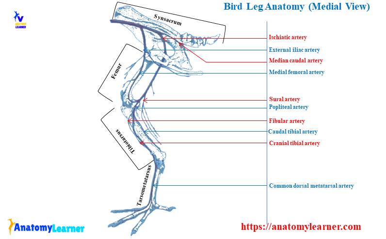
Blood vessels of the bird leg anatomy
The most important blood vessels of the bird leg anatomy are the external iliac artery and ischiadic artery. They arise from the descending aorta of the bird heart. The external iliac artery leaves the descending aorta at the level of the middle renal division. Again, the ischiadic artery leaves the aorta at the level of caudal renal division.
The external iliac artery is smaller than the ischiadic artery in a bird. It passes cranial to the acetabulum and reaches the thigh region of a bird. You will find the following major branches of the bird external iliac artery –
- A pubic artery of a bird and
- The medial femoral artery of the bird.
Again, the medial femoral artery gives the cranial femoral artery and coxal cranial artery. The external iliac artery continues as a medial femoral artery that supplies caudal abdominal muscle and cranial and medial muscle of the bird thigh region.
The ischiadic artery is the largest vessel in the birds leg that passes caudally and dorsal to the hip joint. You will find several branches of the ischiadic artery in the birds leg. At the level of the knee, the ischiadic artery continues as the popliteal artery in a bird.
Then it divides into cranial and caudal tibial arteries at the proximal part of the tibiotarsus bone. The cranial tibial artery of bird continues as the common dorsal metatarsal artery.
Chicken bird leg anatomy labeled diagram
I will show you all the important structures from the chicken bird leg anatomy with a labeled diagram. I tried to show all the important nerves, arteries, and muscles from the chicken leg anatomy. Great, if you want to learn more about the other structures from the leg or other parts of the bird body, you may find your desire articles in the avian anatomy section.
Again, you may follow anatomy learners on social media for more updated leg anatomy labeled diagrams.
Frequently asked questions on birds legs.
Fine, in this part of the article, I will try to solve some common questions on birds legs. If you have any questions related to the birds legs anatomy, please let me know.
What is a bird’s leg called?
The bird’s leg is called the tibiotarsus that is the longest bone in the body. You will also find a slender fibula in the bird’s leg.
Again, in another sense, the bird legs include the femur, tibiotarsus, fibular, and tarsometatarsus bones. Please read the above-mention article, and you will learn the details of the birds’ legs.
What are the leg bones of a bird?
The femur, tibiotarsus, slender fibula, tarsometatarsus, and phalanges are the legs bones. Fine, if you want to know the details anatomy of the leg bones, please read the article.
Do birds have bones in their legs?
Yes, birds have bones in their legs. You will find a total of twenty-one (21) bones in each leg of a bird. The number of leg bones may vary in some breeds of birds.
Do bird legs have muscle?
Yes, bird legs have different muscles that have some clinical importance. The most important muscles from the birds’ legs are iliofubularis, iliotibial, iliotrochanteric, iliofemoral, and femorotibial.
Conclusion
I think the information is enough to learn the bird leg anatomy. But, you might also learn more about the bird leg bones anatomy from another article. It would help if you also learned the bird leg muscle anatomy with the labeled diagram.
For the clinical practice, you might memorize some of the important muscles, course of the sciatic nerve, location of the shank vein from the birds leg anatomy.