The dog spine anatomy consists of vertebrae, intervertebral disc, spinal cord, spinal nerves, and other associated structures. If you are a veterinary practitioner or student, you may treat some common spinal problems of a dog. For that, you might have a good piece of knowledge on the anatomy of a dog spine.
Here, I will show you every structure related to the dog spine with its peculiar anatomical facts. So, you will find detailed guides on dog cervical spine anatomy, lumbar spine anatomy, and more.
I will also show you the differences between the spine of a dog and other mammals. At last, I will try to solve the common questions on dog spine anatomy and injury asked by the owners or veterinary students.
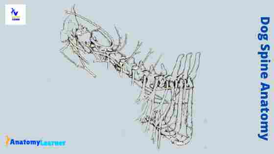
Dog spine anatomy
Fine, first, I would like to summarize the dog spine anatomy. If you find it interesting, you may continue this article to learn the details anatomical facts of dog spine.
So, what should you learn under the spine of a dog? You might focus on the below-mentioned four main structures of the spine –
- Anatomy of the vertebrae from all regions of a dog (cervical, lumbar, thoracic, sacral, and caudal regions)
- Anatomical facts of the intervertebral discs of the dog
- Anatomy of a spinal cord of a dog
- The spinal nerves in different regions of the spine and
- Other associated structures like ligaments, tendons, and muscles are related to the spine.
Let’s know some remarkable anatomical facts of the dog vertebrae –
You will find foramen transversion at the posterior aspect of the atlas of a dog. There is a rod-shaped and pointed odontoid process present in the axis of a dog.
The spinous process of the dog vertebrae is comparatively short compared to the other mammals. But, in the lumbar region, the spinous process becomes broad ventrally and narrow dorsally.
The sacrum of the dog is short and consists of only three vertebrae. You will find well-developed caudal vertebrae in dogs compared to that of an ox or goat.
You will also find some peculiar features in the anatomy of a dog’s intervertebral disc and spinal cord. There are the thickest intervertebral discs present in the cervical and lumbar spine region of a dog.
The length of the spinal cord of a dog is variable according to the breed. You will find thirty-six to thirty-eight pairs of spinal nerves in the dog spine anatomy.
Dog spine anatomy numbers
You might have a question – where the dog’s spine is located? Well, the dog’s spine locates along the dorsal aspect of its body. It starts from the first cervical vertebrae and ends at the caudal vertebrae of a dog.
You will find five different regions of a dog spine – cervical spine, thoracic spine, lumbar spine, sacral spine, and caudal or coccygeal spine. In addition, you will also find seven cervicals, thirteen thoracic, seven lumbar, three sacral, and fifteen to nineteen caudal vertebrae in the spine of a dog.
So, the five divisions of the dog spine are –
- A cervical spine
- The thoracic spine
- A lumbar part of the dog spine
- The sacral part of the spine and
- A caudal or coccygeal part of the dog spine
The number of caudal vertebrae is not fixed in a dog skeleton. You will find a variation in the number of caudal vertebrae in a different breed of dog.
Vertebrae from dog spine anatomy
So, the main part of the dog spine anatomy is the vertebra. Here, I will try to provide all the information on dog vertebrae with the labeled diagram. Ensure you know the name and numbers of vertebrae in the different regions of a dog spine (mentioned earlier).
I think you have a good piece of knowledge on the typical vertebrae of animals. You will find the same structures in the dog vertebrae like body, arch (pedicle, laminae), and different processes as found in an ox or goat.
The adjacent vertebrae of the dog articulate together with the help of ligaments, muscles. And form a vertebral column along the midline of the body. You will also find an intervertebral disc between the adjacent vertebrae of a dog. There are two parts in the dog intervertebral disc – the gel-like nucleus pulposa and annulus fibrosus.
Together with the body, the arch of dog vertebrae forms a short tube (vertebral foramen). All the vertebral foramina concur to form the vertebral canal that contains the spinal cord and roots of the spinal nerves of the dog.
You will find cranial and caudal vertebral notches on either side of the pedicle of a dog vertebra. So, the adjacent vertebrae also form intervertebral foramina. The spinal nerves, arteries, and veins pass through the intervertebral foramina of a dog.
The dorsal part of the arch forms the single spinous process at the middorsal aspect of the dog vertebra. Again, the transverse process projects laterally from the region where the pedicle joins the vertebral body.
In addition, you will find paired articular processes at both the cranial and caudal surfaces of a dog vertebra, at the junction of pedicle and lamina.
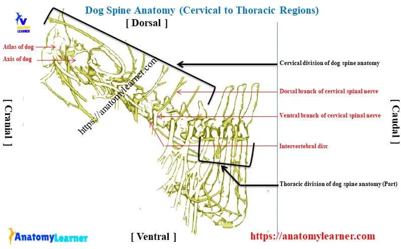
Canine cervical vertebrae anatomy
The cervical part of dog spine anatomy includes the cervical vertebrae, thick intervertebral discs, part of the spinal cord, and cervical spinal nerves. Now, I will describe the anatomical facts of dog cervical vertebrae with a labeled diagram.
In most mammals, you will find seven cervical vertebrae in their vertebral column. You will also find the same number of cervical vertebrae in a dog. The first two cervical vertebrae of a dog (atlas and axis) differing wildly from the others. They can be quickly identified with their peculiar anatomical facts.
But, you will find a bit of anatomical variation on the third, fourth, and fifth cervical vertebrae of a dog. Again, the sixth and seventh cervical vertebrae of dogs possess distinct features that make their identification possible.
Atlas of the dog
The atlas is the first cervical vertebrae in the vertebral column of the dog that is atypical both in structure and function. This vertebra articulates with the skull of the dog cranially and with the axis caudally.
You will find the modified articular processes in the dog that articulates the occipital condyle. There is no spinous process present in the atlas of a dog. The body of the dog atlas is reduced and forms the ventral arch.
The thick lateral mass in the atlas help to unite the ventral arch with the dorsal arch. You will find a shelflike transverse process (wing) that projects from the lateral mass of the atlas vertebrae.
A bifid dorsal tubercle locates on the cranial end of the dorsal arch of the dog atlas vertebra. Again, you will find a ventral tubercle that projects from the caudal end of the ventral arch.
There is a cranial articular fovea present in the dog atlas and consists of two cotyloid cavities. They articulate with the occipital condyles of the dog skull. Again, the caudal articular fovea consists of two shallow glenoid cavities that form a freely movable articulation with the second cervical vertebra.
Fine, a dorsal surface of the ventral arch of the dog atlas possesses a concave fovea dentis that articulate with the long pointed odontoid process of the axis. You will find a sizeable vertebral foramen in the dog atlas that contains the spinal cord.
There are also two pairs of foramina in the atlas of a dog – an alar foramen and a lateral vertebral foramen. The alar foramen is a short canal that passes obliquely through the wing of the atlas. Again, the lateral vertebral foramen locates on the craniodorsal part of the vertebral arch.
Axis of the dog
In the dog spine anatomy, the axis also possesses some peculiar anatomical facts compared to that of the other mammals. You will find a long dorsal spinous process that is bladelike cranially and expanded caudally. The spinous process overhangs the cranial and caudal articular surfaces of the vertebrae.
You will find a cylindrical and pointed peglike eminent (known as the odontoid process) at the cranioventral aspect of the dog axis. The odontoid process of the dog axis is somewhat different morphologically from that of other mammals.
The odontoid process of the dog axis lay on the fovea dentis of the atlas and was held down by the transverse ligament. There is an expanded cranial end on the body of the dog axis that contains the cranial articular surface. Again, the caudal articular processes are a ventrolateral extension of the ventral arch and a spinous process of the axis.
You will find two deep fossae at the ventral surface of the dog axis that separates by the median crest. The cranial vertebral notches concur on either side with those of the atlas from large intervertebral foramina. Through this foramina, the second pair of the cervical spinal nerves pass.
Again, the caudal vertebral notches of the axis help from the intervertebral foramina, passing the third Paris of spinal nerves and spinal vessels.
Other cervical vertebrae anatomy of canine
Okay, the third, fourth, and fifth cervical vertebrae of canine differ slightly from each other. So it is challenging to distinguish these cervical vertebrae of canine perfectly. But the below-mentioned information may help you to identify these cervical vertebrae quickly.
The length of the spinous process of these cervical vertebrae increases from third to fifth. Again, you will find a larger lamine in the third cervical vertebra of a dog. This lamine becomes shorter in the remaining vertebrae of the cervical region.
The caudal articular processes of these cervical vertebrae possess a tubercle. You will find a prominent tubercle on the third cervical, but it gradually decreases.
Again, the transverse process of the fifth cervical vertebrae is short. You will find pairs of transverse foramina in each vertebra (third to fifth).
There is a sizeable spinous process present in the sixth cervical vertebrae of the dog compared to the third, fourth, and fifth cervical vertebrae. But the most peculiar feature of the sixth cervical vertebrae of a dog is the expanded sagittal platelike transverse process (lamina ventralis). You will also find the transverse foramina in the sixth cervical vertebra of a dog.
You will quickly identify the seventh cervical vertebra of a dog. The seventh cervical vertebra of a dog posses the highest spinous process of all those on the cervical vertebrae of a dog.
You will also find costal fovea at the caudoventral aspect of the body of the seventh cervical vertebra of the dog. But, there is no transverse foramen present in the seventh cervical vertebra of a dog.
Thoracic vertebrae in dogs or canine
The thoracic division of dog spine anatomy consists of the thoracic vertebrae, thick intervertebral discs, spinal cord, and thoracic spinal nerves. Now, I will show you some unique anatomical facts of the thoracic vertebrae of a dog so that you may identify it so quickly.
You will find thirteen thoracic vertebrae in the vertebral dog column. The first nine thoracic vertebrae are similar, and the last four contain few variations from each other.
So, what should you know about the thoracic vertebrae of a dog? Well, you should focus on the structure of the vertebral body, arch (pedicle and lamine), and arch that makes it different from other mammals.
Body and arch of the dog thoracic vertebrae
The bodies of the thoracic vertebrae of a dog is shorter than those of the cervical or lumbar region. You will find cranial and caudal costal fovea on either side of the body of the dog’s thoracic vertebrae. But the dog’s eleventh thoracic vertebra posses only the cranial costal fovea.
“The eleventh thoracic vertebra of the dog is known as the anticlinal vertebra.”
The foveae on the bodies of the thoracic vertebrae are for the articulation with the head of a rib. The ventral surface of the most thoracic vertebrae of a dog posses a pair of nutrient foramina. You may also find paired vascular foramina in the dorsal surface of the thoracic vertebrae.
The pedicle of the dog thoracic vertebrae are short than those of the lumbar region. You will find a deep caudal vertebral notch in the pedicle of dog thoracic vertebrae. But, the cranial vertebral notch is absent in the pedicle of thoracic vertebrae.
The lamina is thick and forms the massive and long spinous process (most characteristics feature of thoracic vertebrae).
The spinous process of the dog thoracic vertebrae
The massive and long spinous process is the most identifying anatomical feature of the dog thoracic vertebrae. You will find a more massive spinous process in the first thoracic vertebra of a dog. The massiveness of the spinous process gradually decreases with the successive vertebrae.
There is a minor change in the length and direction of the dog thoracic spinous process until the seventh and eighth vertebrae. You will find a short and caudally inclined spinous process in the nine and tenth thoracic vertebrae.
Again, the spinous process of the dog eleventh thoracic vertebrae is nearly perpendicular to the long axis of the body. In addition, the spinous processes of the first tenth thoracic vertebrae direct caudally. But, from the eleventh to last thoracic spinous processes, they directed cranially.
Another process of the dog thoracic vertebrae
Great, it would help if you also learned the anatomical facts of the dog thoracic vertebrae’s transverse, articular, and accessory processes. The transverse processes are short, blunt, and irregular in dog thoracic vertebrae.
You will find foveae in all the transverse processes of the dog thoracic vertebrae for articulation with the tubercle of a rib. The size and convexity of these foveae decrease gradually from first to last thoracic vertebrae.
Sometimes, you may find the slight, knoblike eminence that projects dorsally from the transverse process of the second or third thoracic vertebrae. At the eleventh thoracic vertebrae level, they become associate with the cranial articular processes and continue as laterally compressed tubercles throughout the remaining vertebrae.
The articular processes of the dog thoracic vertebrae are located at the junction of the pedicle and laminae. You will find a widely separated cranial articular process in the first and second thoracic vertebrae. Again, in the third to tenth thoracic vertebrae of a dog, the cranial articular processes are nearly confluent at the median plane. But, the articular processes of the eleventh, twelfth, and thirteen thoracic vertebrae face each other across the median plane.
The cranial articular process of the dog thoracic vertebrae fac cranially and dorsally, except the last three. In addition, the caudal articular process is somewhat modified in the dog thoracic vertebrae.
You will find a well-developed accessory articular process the extend caudally from the caudal border of the pedicle. The accessory process also forms a notch with the caudal articular process of a dog thoracic vertebrae.
Dog lumbar spine anatomy
The lumbar vertebrae are an essential component in the lumber division of dog spine anatomy. You will find seven lumbar vertebrae in the vertebral column of a dog. Let’s know the unique osteological features of the dog lumbar vertebrae.
The lumbar vertebrae of a dog possess a longer body than that of the thoracic vertebrae. They gradually increase the length from the first to fifth lumbar vertebrae. The length of the first and seventh lumbar vertebrae of the dog is almost the same.
In addition, the body of the dog lumbar vertebrae is circular in appearance compared to that of the ruminant. The width of the body gradually increases throughout the series of lumbar vertebrae.
You may find single or paired ventral foramina at the ventral surface of the lumbar vertebrae body. But, sometimes, some lumbar vertebrae of the dog lack these ventral foramina on the ventral surface.
Again, you will find the paired dorsal foramina on the dorsal surface of the body of lumbar vertebrae. The pedicle and lamina of the dog lumbar vertebrae are more massive and more prolonged. They possess a typical osteological feature, as you found in other regions of the vertebrae.
The processes of the dog lumbar vertebrae
You will find longer transverse processes in the dog lumbar vertebrae, the most identifying characteristics features. The transverse process of the dog lumbar vertebrae directs cranially and slightly ventrally. These features are also unique compare to that of ruminant lumbar vertebrae.
Again, the transverse process of the dog lumbar vertebrae is most prolonged at the mid lumbar region of the skeleton. You may easily palpate the transverse processes of the dog’s lumbar vertebrae.
The length of the spinous processes of the lumbar vertebrae is less than that of the dog thoracic vertebrae. But, the dorsal border of the lumbar spinous processes is wider, two times higher than that of the thoracic spinous process.
You will find the highest and more massive spinous processes in the mid lumbar region of a dog skeleton. The spinous process of the dog lumbar vertebrae has a slight cranial inclination.
The articular process of dog lumbar vertebrae lies mainly in the sagittal plane. You will find the caudal articular process of the dog lumbar vertebrae in between the cranial articular processes of the succeeding vertebrae. In addition, all the cranial articular processes of the dog lumbar vertebrae possess a mammailry process.
You will find well-developed accessory processes on the first three or four lumbar vertebrae of a dog. But, there are no accessory processes on the fifth or sixth lumbar vertebrae of a dog. The accessory processes overlie the caudal vertebral notches and extend caudally lateral to the articular process of the succeeding vertebrae.
Sacral vertebrae of dog (sacrum)
In the sacral division of the dog spine anatomy, the vertebrae are an essential component. You will find three sacral vertebrae that fuse in older dogs and forming the sacrum.
The bodies and arches of these three sacral vertebrae fuse from the arch, bony mass – the sacrum. It is a four-sided and wedge-shaped structure that lies in between the ilium bone of the dog.
The body of the first sacral vertebra of a dog is larger than the bodies of the other two vertebrae. You will find an enlarged wing, a base, a concave ventral surface, and a convex dorsal surface in the dog sacrum.
Base, apex, and wing of the dog sacrum
You will find a convex base of the dog sacrum body that faces cranially. The three sacral foramina form the sacral canal, in which the sacral part of the spinal cord passes. There is a promontory that finds at the cranioventral part of the base. This is a transverse ridge at the ventrolateral aspect of the base.
This structure also helps to form the small part of the pelvic inlet in a dog. The apex of the sacrum is the caudal extremity of the dog sacrum. It is broad transversely and articulates with the first caudal vertebra of the dog.
“Sometime, you may find the fusion of first caudal vertebrae with the sacrum in some breed of the dog.”
The craniolateral part of the dog sacrum enlarges and forms the wing. You will find large and rough articular surfaces in the wing that articulates with the ilium bone.
Dorsal surface of the dog sacrum
The dorsal surface of the dog sacrum is slightly convex and possesses some essential structures. You might identify the flowing structures from the dorsal surface of the dog sacrum.
The cranial articular processes
A median sacral crest of the dog sacrum bone
The caudal articular processes
Four dorsal scaral foramina of the dog sacrum
Two intermediate sacral crests and
The lateral sacral crest of the dog sacrum bone
You will find a median sacral crest on the dorsal surface of the dog sacrum. This median sacral crest forms by the fusion of the spinous processes of three sacral vertebrae.
You will also find the intermediate and lateral sacral crest on the dorsal surface of the dog sacrum. There is an extensive cranial articular process present at the dorsomedial aspect of the dog sacrum. It joins with the caudal articular process of the last lumbar vertebra.
Again, there is a small caudal articular process find in the dog sacrum. It articulates with the first caudal vertebra of the dog.
In addition, you will find four dorsal sacral formations on the dorsal surface of the dog sacrum. The dorsal division of the sacral spinal nerve and spinal vessels are passing through these foramina.
Caudal vertebrae and spine of the dog anatomy
The caudal vertebrae and spine of the dog anatomy have a significant variation than the other parts. You will find fifteen to nineteen caudal vertebrae in the vertebral dog column. But, this number of the caudal vertebrae may vary with the different breeds of dogs.
You may find the body, arch, and processes in the first few caudal vertebrae of a dog skeleton. But, the last caudal vertebrae lose their distinct features and become simple rods.
The body and arch of the dog caudal vertebrae
The body of the first caudal vertebrae is as wide as it is long. Again, the length becomes gradually increases up to the middle segment of the caudal vertebrae. But, in the last few caudal vertebrae, you will find the shorter length.
The width of the dog caudal vertebrae gradually decreases in the series. You will find a tapering process at the end of the last caudal vertebra of a dog.
In the first caudal vertebrae, you will find a well-developed arch. But the other caudal vertebrae possess atypical arches, and the last few lose them. There is a small caudal vertebral canal present in the vertebral dog column. The caudal part of the spinal cord passes through this small canal of the caudal vertebrae.
The processes of the dog caudal vertebrae
The processes of the dog caudal vertebrae are not so developed as found in the other part of the vertebral column. You will find a small spinous process in the dog caudal vertebrae. But this spinous process disappears in the last segment of this series.
You may find a well-developed and typical transverse process in the first few caudal vertebrae. These reduce in size in the middle segments and disappear in the last few segments of caudal vertebrae.
You will also find the cranial articulation processes in the dog caudal vertebrae. But, some of these cranial articulation loses their articular function in the last segments. There may present the mammillary process in the cranial articular process of the dog caudal vertebrae.
The caudal articular processes are short in the dog caudal vertebrae. They project from the lateral border of the arch and are frequently asymmetric. Again, they disappear in the last few caudal vertebrae of the dog.
Joints of the vertebral column of a dog
You will find the same type of joints in the vertebral column as found in the ruminant. Here, I will provide a little information about the joints of the vertebral column of a dog.
Generally, you will find the below-mentioned joints in the vertebral dog column –
- Atantooccipital joint of a dog
- An atlantoaxial joint of the vertebral dog column
- The intercentral joint of the dog vertebrae
- Intraneural joints of the dog vertebrae
- Intertransverse joints of the dog vertebrae
- The lumbosacral joint of the vertebral dog column
- Sacrococcygeal joints of the vertebral dog column
- The intercoccygeal or caudal joints in the vertebral dog column
The involvement of the bones and ligaments in forming the joints mentioned above are the same as found in the ruminant vertebral column.
The atlantooccipital joint of the dog vertebral column forms by the dorsolaterally extending occipital condyle and the corresponding concave cranial articular fovea of the atlas. Again, the atlantoaxial join of the dog vertebral column forms by the fovea dentis of the atlas and the odontoid process of the axis.
In addition, the intercentral joint of the dog vertebrae forms by the caudal concave surface of the receding vertebrae and convex cranial surface of the preceding vertebrae. The interneural joints of the dog vertebrae form by the arch and pedicle of the vertebrae.
The lumbosacral joint of the dog vertebral column forms between the last lumbar vertebrae and the first sacral vertebrae or sacrum.
Intervertebral discs of the dog vertebrae and spine anatomy
The intervertebral discs are another main component of the vertebral dog column and spine anatomy. These intervertebral discs are interposed in every intervertebral space and uniting the bodies of the adjacent vertebrae.
The thickness of the intervertebral discs is excellent in the cervical and thoracic region of the vertebral dog column. Again, you will find the thinnest intervertebral discs in the caudal vertebrae of the dog.
“The intervertebral spaces between first two or three cervical vertebrae possess the thickest intervertebral discs.”
Grossly, the dog intervertebral discs possess two defined portions –
- An outer laminated fibrous ring (annulus fibrosus), and
- A central, amorphous, and gelatinous nucleus pulposus.
Fibrous annular and nucleus pulposa parts of the dog intervertebral disc
The fibrous annular part of the dog intervertebral disc consists of a brand of parallel fiber that runs obliquely. In addition, the ventral part of the annulus fibrossus is three-time thicker than the dorsal part. The band of the fiber crosses each other and forms the latticelike pattern.
They provide the transmission of stresses and strains that requires by all the lateral and dorsoventral movement.
The nucleus pulposa of the dog intervertebral disc is the remnant of the notochord. It is a depressed area on each end of the vertebral body surrounded by the fibrous ring. The consistency of this nucleus pulposa is semifluid, and it is put under pressure by any movement of the vertebral bodies.
Some ligaments of the dog vertebrae and spine anatomy
I am not going to details the description of the ligament in the dog vertebral articulation and spine anatomy. Here, you will find a list of ligaments that are associated with the vertebral joints of the vertebral dog column.
A lateral ligament of the atlantooccipital joints – runs from the lateral part of the dorsal arch of the atlas to the paracondylar process of the occipital bone.
An apical ligament of the dens – it leaves the apex of the dens and passes straight cranially to the basioccipital bone.
The two alar ligaments of the dog atlas,
The transverse Atlanta ligament – is a thick ligament that connects one side of the ventral arch of the atlas to the other.
You will find some long ligaments in the vertebral dog column. The long ligaments of the vertebral dog column are –
The nuchal ligament extends caudally to the dorsal extremity of the spinous process of the first thoracic vertebrae.
A supraspinous ligament is another long ligament that extends from the spinous process of the first thoracic vertebrae to a dog’s third caudal vertebrae.
The ventral longitudinal ligament – lies on the ventral surface of the body of dog vertebrae,
A dorsal longitudinal ligament – lies on the dorsal surface of the body of the dog vertebrae.
You will also find the inter capital ligament, interspinous ligament, intertransverse ligament, yellow elastic ligament, and ligaments for the ribs head in the vertebral dog column.
Spinal cord from dog spine anatomy
Now, let’s learn the essential features of the spinal cord from the dog spine anatomy. The spinal cord of a dog is enclosed within the vertebral canal. It covers by thick meninges that have three defined portions – dura mater, pia mater, and arachoined mater. You may learn the details of gross anatomy and histology of the animal spinal cord from the other article of anatomylearner.com.
In the dog spinal cord, you will find the dorsal and ventral roots of the spinal nerves that cover the meninges. As I told you before, the meninges of the dog spinal cord has three layers; the first and superficial layer is the dura mater which is thick and fibrous.
There is a thin arachnoid matter that lines the inner surface of the dura mater. Again, you will find the thinnest vascular pia mater on the spinal cord surface.
The dog spinal cord comprises the following structures –
- A central canal of the dog spinal cord,
- The gray matter of the dog spinal cord, and
- A white matter of the dog spinal cord
The central canal lies on the center of the spinal cord and contains cerebrospinal fluid. Again, the gray matter of the spinal cord consists of cell bodies, processes of neurons, and glial cells. It is a butterfly-shaped bilateral wing connected at the midline by the central intermediate substances.
You will also find a lateral intermediate substance known as the lateral horn of the dog’s spinal cord. Dorsal to the lateral intermediate substance, there is a dorsal horn present in the spinal cord. You will also find a ventral horn, ventral to the lateral intermediate substance.
The white mater of the dog’s spinal cord locates superficial and contains the myelinated axons (Labeled diagram).
The segment of the dog spinal cord
At the bird, the dog spinal cord extends into the sacrum vertebrae. But the following postnatal development, the dog spinal cord terminates in the caudal region of the lumbar vertebrae. So, you will find a well-developed segment of the spinal cord up to the last lumbar vertebrae.
The most extended spinal cord segment of the dog is found in the cervical region. Again, the dog spinal cord’s thoracic segment is also more significant compared to the lumbar part.
Caudal to the dog lumbar vertebrae, spinal cord tapers into an elongated cone (Conus medullaries). You will find some terminal filament at the end of the Conus medullaries. Collectively, these terminal filaments are known as the cauda equine.
In the dog, most of the caudal equine lies caudal to the lumbar vertebral canal.
Dog spinal nerve anatomy
You will find the thirty-six to thirty-eight pairs of spinal nerves in the vertebral dog column. I am not going to details the anatomy of these spinal nerves from the dog. But, you might have a good piece of knowledge on the structure of a spinal nerve and the list of spinal nerves from dog anatomy.
You will find the following general features in the dog spinal nerve –
The root segments – a dorsal and ventral roots; a spinal ganglion,
Four primary branches segments – dorsal, ventral, meningeal, and communicating branches of spinal nerve
The peripheral branch segments – a dorsal and ventral peripheral branch of dog spinal nerve
I hope the dog spinal nerve anatomy diagram might help you to understand all these branches so easily.
You will find the spinal nerve in the cervical, thoracic, lumbar, sacral, and caudal regions of the dog anatomy. There are eight pairs of cervical spinal nerves present in the dog. Again, you will find thirteen Paris of a thoracic spinal nerve in the dog.
The lumbar spinal nerves of a dog are seven in number on each side. In addition, you will find three sets of dorsal and ventral roots that merge to form the sacral spinal nerve in the dog.
The number of caudal spinal may vary in a different breeds of dogs. But, you may find three to six caudal spinal nerves in the most dog breed.
Frequently asked questions on dog spine.
Fine, here you will find the answers to frequently asked questions on dog spine. If you think this information is not enough, feel free to ask me any questions related to dog spine anatomy.
How do you know if your dog has spinal problems?
You may find different problems in your dog if it has spinal pain. The abnormal posture, lower head carriage, shivering, rounding of the back region, refusal to get up and play are most common if your dog has a spinal problem.
What is the weakest area of a dog’s spine?
The weakest area of a dog’s spine is the cervical region. In this cervical region, you will find seven cervical vertebrae. The intervertebral disc of this area is generally thickest. Again, the spinal cord is also more significant and typical in the cervical region of dog anatomy.
You may also find the weak area in the lumbar spine region of a dog.
How many vertebrae do dogs have?
The number of dog vertebrae may vary in the different breeds. But usually, you will get seven cervicals, thirteen thoracics, seven lumbar, three sacral vertebrae, and fifteen to nineteen caudal vertebrae in the dog skeleton. So, the total number of vertebrae maybe forty-five to fifty (45 – 50) in a dog skeletal system
What is a herniated disc in a dog?
Why can I see my dog’s spine?
It is normal to feel the dog’s spine externally. This is because there is a thin layer of fat under the skin of a dog.
Is it normal to feel the spine of a dog?
Yes, it is normal to feel a dog’s spine. As there is a thin layer of fat under the skin, you may feel the dog’s spine by external palpation.
Can a dog survive spinal injury?
Yes, can survive after a spinal injury. But the time of recovery may vary with the condition of the spinal injuries in a dog.
Where is the cervical spine in a dog?
Where does the spinal cord end in a dog?
In a young dog, the spinal cord ends at the sacral vertebrae. But in older dogs the spinal cord ends at the last lumbar vertebrae. At the end of the lumbar vertebrae, the spinal cord forms the Conus medullaries. In addition, you will find the terminal filament in the caudal vertebral region of the dog. All these terminal filaments collectively form the cauda equine in the dog tail.
How many vertebrae are in dog tail?
The tail of the dog comprises caudal vertebrae. But, the number of caudal vertebrae is not fixed in the dog tail. You may get fifteen to nineteen vertebrae in the tail of a dog.
How many caudal vertebrae do dogs have?
The caudal vertebrae of the vertebral dog column may vary in the different breeds. But, generally, you will find fifteen to nineteen caudal vertebrae in the vertebral dog column. Typical osteological features are not found in these caudal vertebrae of the dog.
Conclusion
So, you might know the detailed structure of dog vertebrae, intervertebral discs, spinal cord, and spinal nerve if you want to get complete knowledge on dog spine anatomy. Here, I only focus on the dog vertebrae anatomy, but you may learn the detailed anatomical facts of the spinal cord and spinal nerves from another related article.
You might have a good piece of knowledge on the dog cervical spine anatomy and dog thoracic spine anatomy, as most of the spinal injuries occur in these regions.