Near the junction of the oral cavity and the pharynx, you will find several collections of lymphoid tissue that refer to the tonsil. In tonsil histology, different essential structures like tonsillar crypts, lining epithelium, numerous lymphatic nodules, and dense connective tissue capsules should be identified under a light microscope.
I will show you all these structures from the palatine tonsil histology slide. Here, I will provide the identifying points for the palatine tonsil slide with a concise description. Again, you will find a little information on the other different tonsils like pharyngeal, lingual, and tubal tonsils with the labeled diagrams.
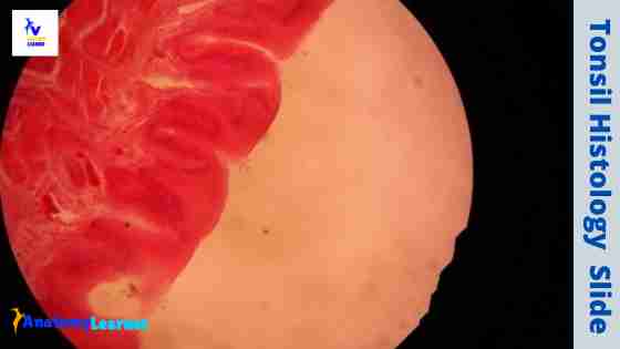
Tonsil histology
The tonsil is located adjacent to the host organs’ lumen that is covered with either nonkeratinized stratified squamous or pseudostratified columnar epithelium. There are four different types of tonsils in the animal – palatine, pharyngeal, lingual, and tubal.
The two most prominent (right and left) palatine tonsils are present on either side of the oropharyngeal isthmus. The histology of palatine tonsil consists of aggregated lymphatic nodules, tonsillar crypts that cover with the nonkeratinized stratified squamous epithelium.
You will also find the collection of lymphatic nodules on the posterior wall of the pharynx. This is known as the pharyngeal tonsil that also possesses the same characteristics features as the palatine tonsil.
Again, in the dorsum of the posterior part of the tongue, there is a smaller collection of lymphatic nodules. This is the lingual tonsil that I will show you later in this article. In addition, you will find the tubal tonsil around the pharyngeal openings of the auditory tubes.
“In this article, I will only represent the details guide on palatine tonsil.”
What should you do if you are asking to identify the palatine tonsil under the light microscope at your laboratory? You should identify the following essential structures from the palatine tonsil histology slide.
- A nonkeratinized stratified squamous epithelium lining
- The deep groove or tonsillar crypts of the tonsil
- Numerous lymphatic nodules along the length of the tonsillar crypts
- The trabeculae with blood vessels
- Germinal centers of the lymphatic nodules
- A dense connective tissue capsule of the tonsil
That’s fine; now, let’s try to find out these structures from palatine tonsil slides.
Palatine tonsil histology slide identification points
The following essential identification points might help you quickly identify the palatine tonsil histology slide. If you want, you may add more identification points for identifying palatine tonsil slides under a light microscope.
- The sample tissue section lines with the nonkeratinized stratified squamous epithelium
- Presence of tonsillar crypts (deep grooves) that also lines with the nonkeratinized stratified squamous epithelium
- The tissue section shows numerous lymphatic nodules that distribute along the lengths of tonsillar crypts.
- There is a dense connective tissue capsule that underlies the sample tissue section.
So, this is a palatine tonsil histology slide.
Palatine tonsil histology description
The right and left palatine tonsils consist of aggregates of lymphatic nodules located in the oral cavity of an animal. You will find a variation in the surface of the palatine tonsil in different species. The surface may be relatively smooth in the case of a dog and cat’s palatine tonsil.
But, the palatine tonsil of ruminant and horse possess deep tonsillar crypts. The lingual tonsil of the horse also contains the deep grooves or tonsillar crypts.
The surface of the palatine tonsil is covered by the nonkeratinized stratified squamous epithelium that also covers the rest of the oral cavity. You may also find the pseudostratified columnar epithelium on the surface of some palatine tonsil histology.
Below the lining epithelium (nonkeratinized stratified squamous epithelium), you will find the collection of numerous lymphatic nodules. These lymphatic nodules are distributed along the entire length of the tonsillar crypts (deep grooves of the tonsil structure).
The lymphatic nodules frequently merge. You will find the deep-staining zone in the lymphatic nodules of the tonsil slide. Again, there is a light-staining part in each lymphatic nodules of the palatine tonsil. The lymphatic nodules’ lighter-staining parts are known as the germinal center.
You will find a dense connective tissue capsule that underlies the palatine tonsil structure. This thick connective tissue capsule forms the trabeculae that pass towards the tonsil surface between the lymphatic nodules.
You will find numerous blood vessels in the trabeculae of the tonsil. Sometimes, many mucosal glands may present in the deeper part of the tonsil just outside of the capsule.
The ducts of the mucosal glands open at the bottom of the crypts. You will also find the skeletal muscle fibers under the connective tissue capsule of the tonsil.
Other information on palatine tonsil
The tonsillar crypts of the palatine tonsil allow a high concentration of the lymphatic tissue in a given area. Again, the epithelium of the palatine tonsil usually infiltrates with the variable degree of lymphocytes, neutrophils, and macrophages.
This infiltration is particularly more in the tonsil of the oropharynx. In addition, beneath the epithelium, diffuse lymphatic tissue with plasma cells surround the lymphatic nodules.
The tonsil belongs to the mucosa-associated lymphatic tissue (MALT). But, as a dense connective tissue capsule partially covers the tonsil, it is considered an organ.
If you want to learn about mucosa-associated lymphatic tissue (MALT), please read the following part of the article.
Mucosa-associated lymphatic tissue (MALT)
The significant aggregation of lymphocytes may be found in the mucosa of an animal’s respiratory, alimentary, and urogenital tracts. These aggregated lymphatic nodules are termed the mucosa-associated lymphatic tissue.
The total volume of the mucosa-associated lymphatic tissue is more or less equal to that of the lymphoid tissue present in the lymph node and spleen. These mucosal lymphatic tissue functions as an integrated mucosal immune system to augment the chemical and mechanical barriers of the surface epithelium.
You will find solitary lymphatic nodules, as well as aggregated lymphatic nodules under the lining epithelium of the most mucosa membrane. You will find the following common characteristics features in most mucosal-associated lymphatic tissue.
The lymphocytes aggregate in a single or more follicle or nodules. You will find a similar structure in the lymph node. The diffuse lymphatic tissue may present in the interval between the lymphatic nodules. You will also find a lighter-stained germinal center in each nodule of the structure.
The lymphatic nodules of the mucosa-associated lymphatic tissue predominantly contain the B-Lymphocytes, whereas the diffuse area has T-Lymphocytes. The lymphoid nodules present very close to the lining epithelium of the organs.
If there is a larger aggregation of lymphatic nodules, it may extend up to the tunica submucosa layer. Again, the individual lymphocyte may infiltrate the epithelium and pass through it into the lumen of the specific organs.
You will not find any connective tissue capsule that covers these lymphatic nodules. But a supportive network of reticular fibers and a dense connective tissue capsule may present beneath the lymphatic nodules.
Types of mucosa-associated lymphatic tissue
You will find a small aggregation of lymphatic nodules in the respiratory tracts (especially in the trachea wall and larger bronchi). This small aggregation of lymphatic nodules and tissue may be defined as the bronchial associated lymphoid tissue (BALT).
Again, you will find a large aggregation of lymphatic tissue and nodules in the alimentary canal of an animal. More lymphatic nodules may discover in the end part of the small intestine of an animal. These more significant lymphatic nodules and tissue aggregation are formally known as the gut-associated lymphoid tissue. The best example of gut-associated lymphoid tissue is tonsil, Peyer’s patches, and lymphatic nodules in the vermiform appendix.
Anatomy of palatine tonsil
In this part, I will share a little information on the anatomy of a palatine tonsil from an animal. It is an almond-shaped structure located on the oropharynx’s lateral wall in the tonsillar fossa. Again, the palatine tonsil bounds by the palatoglossal arch in front and palatopharyngeal arch behind.
The lateral surface of the palatine tonsil lies on the tonsillar bed that covers the dense connective tissue capsule. Again, the medial surface of the palatine tonsil shows different orifices that lead into crypts inside the tonsilar substance.
Pharyngeal tonsil histology
The pharyngeal tonsil is present on the posterior wall of the pharynx. You will not find any defined tonsillar crypts in the pharyngeal tonsil histology slide. The lining epithelium of the pharyngeal tonsil is pseudostratified columnar epithelium (ciliated).
The epithelium shows a small fold, and the lamina propria contains diffuse lymphatic tissue and lymphatic nodules. In addition, the capsule underlies the lymphatic nodules that are comparatively thinner than that of the palatine tonsil.
Lingual and tubal tonsil
You will find the lingual tonsil on the dorsum of the posterior part of an animal’s tongue. The small and rounded aggregations of lymphatic nodules greatly vary with shape and size. The single tonsillar crypts are defined in the lingual tonsil histology slide. The surface of the lingual tonsil lines with the nonkeratinized stratified squamous epithelium.
Again, you will find the tubal tonsil around the pharyngeal opening of the auditory tube. There are small lymphatic nodules found in the lamina propria of the tubal tonsil. The tonsillar crypts are not so well developed in the tubal tonsil slide.
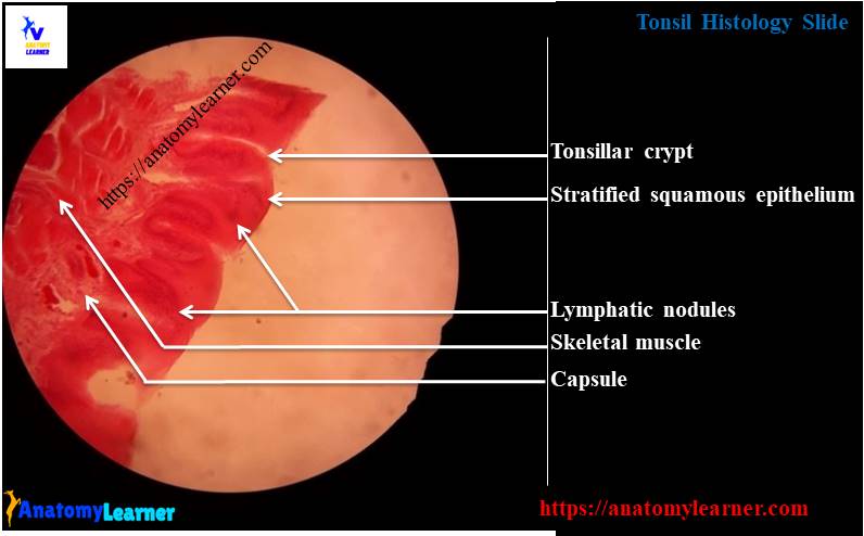
Palatine tonsil histology slide labeled diagram
Finally, I will show you all the palatine tonsil histology slide structures with the labeled diagram.
Here, I tried to show you all the histological features of a typical tonsil structure. So, you may also use this diagram for other tonsils (with minor modification).
Here, the surface of the palatine tonsil covers the nonkeratinized stratified squamous epithelium. The tonsillar surface form the deep grooves (known as the tonsillar crypts). These tonsillar crypts also line with the nonkeratinized stratified squamous epithelium (shown in the diagram).
The diagram also shows the numerous lymphatic nodules throughout the length of the tonsillar crypts. Here, I also marked the deep zone and the lighter zone of the lymphatic nodules. The lighter area of all lymphatic nodules in the germinal center (shown in the diagram).
Again, the labeled diagram also shows some diffuse lymphatic nodules present in the tonsil structure. A dense connective tissue capsule is also marked in the labeled diagram underlies the palatine tonsil. I also tried to show some of the connective tissue trabeculae that contain numerous blood vessels.
The palatine tonsil labeled diagram shows some skeletal muscle fibers under the connective tissue capsule. But this diagram does not show the mucosal gland just under the lymphatic nodules.
For your kind information, I will try to update the tonsil histology slide in the future. You may join anatomy learner on social media if you want to get more tonsil microscope slide images and labeled diagrams.
Frequently asked questions on tonsil slides.
So, in this part of the article, I will try to solve the common inquiries on the tonsil histology slides. I hope you find your desired answers to the following questions on tonsil histology. These questions on tonsil might help your viva-voce.
What type of epithelium is in the tonsils?
Fine, if you read the article from the beginning, you will know about the tonsil’s epithelium. However, you will find two different types of lining epithelium on the tonsil surface. In most animal palatine tonsils, you will find the nonkeratinized stratified squamous epithelium. Again, there is a pseudostratified columnar epithelium lining in the animal’s palatine tonsils.
In the pharyngeal tonsil of animals, you will find the pseudostratified columnar ciliated epithelium lining. Again, in the lingual and tubal tonsil, the lining epithelium is the nonkeratinized stratified squamous epithelium.
I hope you have a good piece of knowledge on the histology of nonkeratinized stratified squamous epithelium and pseudostratified columnar epithelium. If you want to memorize them, you may read this article from anatomy learner.
The stratified squamous consists of many layers of cells (columnar, polyhedral, and squamous). There are flat cells (squamous) with elliptical nuclei present in the superficial layer of the stratified squamous.
The pseudostratified columnar epithelium consists of cells of different shapes and heights lying on the basement membrane. You will find a hair-like process (cilia) on the free surface of the epithelium.
What is the structure of the tonsil?
I have already described all the structures of the tonsil histology slide with diagrams. You may read all the information, but now I will provide a short note on tonsil structures. You will find a significant aggregation of lymphatic tissue and nodules that are readily recognized because it is covered by stratified squamous epithelium (nonkeratinized). The lining epithelium of the structure dips into the tonsil to form the deep groove (tonsillar crypts).
There are numerous lymphatic nodules on the whole length of that tonsillar crypts. You will find a germinal center in each lymphatic nodules of the tonsil structure. The lymphatic nodules may extend into the submucosal layer in some tonsil structures.
In the tunica submucosa layer of the tonsil structure, you will find the mucosal glands (but not occur spontaneously). A capsule does not cover the lymphatic nodules of the tonsil, but you will find a dense connective tissue capsule under the lymphatic nodules.
Again, the microscope slide of the tonsil shows skeletal muscle fiber under the dense connective tissue capsule.
What are the 4 types of tonsils?
The aggregated lymphatic nodules and diffuse lymphatic tissue near the larynx and oral cavity are known as the tonsil. You will find four different types of tonsils in animals – palatine tonsil, lingual tonsil, pharyngeal tonsil, and tubal tonsils.
If you want to learn the histology of palatine tonsil with the labeled diagram, you may read the above information. I hope you will learn the basics of the histological features of a tonsil.
What tissues are found in the tonsils?
You will find the lining epithelium, connective tissue framework, diffuse lymphatic tissue, and lymphatic nodules in the tonsil structure. In this article, I have already described the types of lining epithelium (nonkeratinized stratified squamous and pseudostratified columnar epithelium). Again, you will find the lamina propria and tunica mucosa that contains connective tissue and glands.
The framework of the tonsil forms by the connective tissue reticular fibers. Again, the essential tissue in the tonsil structure is aggregated lymphatic tissue or nodules (primarily B-lymphocytes). In addition, you will also find the muscular tissue (skeletal muscle fibers) in the tonsil structure.
If you want to konw more about the issues found in the tonsil, please reread the full article. Again, you may know the histology of the following lymphoid organs from the animal body –
- Histological features of lymph node with slide images and labeled diagrams
- Thymus gland histology slide and labeled diagrams
What is connective tissue is in the tonsils?
Conclusion
I think you got the basic idea on the tonsil histology slide. All the labeled diagrams of the palatine tonsil histology slide might help you identify it under the light microscope. It would be best to make a difference among the histology of the different types of tonsils – palatine, pharyngeal, lingual, and tubal tonsils.
Again, it would help to identify the tonsil histology slide under the light microscope with the help of identification points that I provided in this article.