There are 4 paws in a cat that contains 18 claws altogether. In the cat paw anatomy, you will learn the bones, muscles, vessels, and digital pad structures in detail. I will show you all the cat paw pad anatomy structures with a diagram from both front and hind legs.
Again, I will show you the difference between the normal cat and the polydactyl condition with a possible explanation. Again, you will find your answers on the carpal and metacarpal pads of the cats in detail.
So, if you are interested to know the meaning of cat paw, its structures, and variation in a different breed of cat, you may continue this article till the end.
Cat paw anatomy
The cat paw anatomy includes the bones, muscles, vessels, and other surrounding structures from the regions of manus and pes. You will find 4 paws altogether in cat legs which are structurally almost similar. The exceptional features of the cat’s front and hind paw will be discussed in the specific part of the article.
First know, what the cat paws are? Well, I would like to answer this question in a very simple way so that you may understand the cat’s paws easily. The cat’s paw resembles the human hand. So, the cat’s front paw includes the carpal, metacarpal, and phalanges regions.
Again, the cat’s hind paws include tarsal, metatarsal, and phalanges regions along with their anatomical features. At the end of each 4 paws of the cat, you will find 4 – 5 toes or claws.
Usually, there are 5 toes (claws) present in each forepaw of the cat (4 developed and 1 dewclaw). Again, the hind paws of the cat contain 4 developed claws. But, you may find more or fewer claws in both the front and hind paws of the cats.
From both the front and hind paws of the cat, you might learn the anatomical features of bones, muscles, vessels, and nerves. Let’s get a basic idea of the forepaws and hind paws of the cat in a little.
Features of the cat forepaw
The bones or skeleton of the cat’s front paw includes the carpus, metacarpus, phalanges, and special sesamoid bones associated with them. There are seven carpal bones in the cat front paw that arranges into two rows. You will find five metacarpal bones in the cat front paw anatomy; these are articulates with carpal bone proximally and phalanges distally.
So, you could understand that there are five digits (toes) present in the cat’s front paw. And each of these toes is formed by the phalanges.
The first toes of the cat consist of only two phalanges, and the other four toes contain three phalanges. You will also find some sesamoid bones in the metacarpal and phalanges. I will discuss this in detail in the paws bones anatomy section.
Several muscles run over the carpus, metacarpus, and supply to the cat’s digits. The most important muscles of the cat front paws are extensor digitorum communis, extensor digitorum lateralis, flexor digitorum profundus, flexor digitorum superficialis, and lumbrical muscles.
These muscles form the strong ligaments and tendons, important structures in the cat’s front paw. The cranial superficial antebrachial branches of the radial, median and ulnar arteries supply the front paw of the cats.
Again, the radial, median, and ulnar nerves branch from the dorsal and palmar common digital nerves. These common digital nerves again divide into proper dorsal and palmar nerves that supply to the cat’s front paw.
You will also find some other important structures in the front paw of a cat. Other important structures of the front paw of the cats are the carpal pad, metacarpal pad, and digital pad.
Features of the cat hind paw
You will find almost similar structures in the hind paw of a cat, like a front paw. The bones of the hind paw of a cat include the tarsus, metatarsal, phalanges, and special sesamoid bones. You will find 7 tarsal bones in the hind paw of the cat that arranges in two transverse rows.
These tarsal bones articulate with the four or five metatarsal bones at their distal ends. Each of the metatarsal bones of the cats hind paw contains three phalanges and sesamoid bones. They form the four developed digits in the hind paw of the cats.
The first digit is usually absent in the cat’s hind paw. But, sometimes, you may find a rudimentary digit (first) in some cat breeds.
Some important muscles run over the tarsal metatarsal and supply to the cat’s hind paw digit. The most important muscles of the cats hind paw are extensor digitorum longus, brevis, flexor digitorum longus, brevis, minimal digital abductor, and lumbrical muscles.
These muscles also form the short digital ligaments and tendon in the hind paw of the cat. The branches of cranial tibial, cranial, and caudal medial saphenous arteries supply the cat’s hind paw. Again, you will find the branches of the cranial tibia and medial saphenous vein in the hind paw structure of the cats.
The sciatic nerve of the cats gives several branches where the tibia and fibula nerve supply up to the hind paws. Both the deep and superficial tibia nerves give the dorsal common digital nerves supply to the dorsal aspect of the hind paw. Again, the branches of the tibia nerves supply the lateral and plantar aspects of the hind paws.
You will find the digital pads and metatarsal pad in the hind paw. But, there is no tarsal pad present in the cats.
Cat front paw anatomy
So, in the cat front paw anatomy, I will discuss the bones, muscles, vessels, and nerves. I will also describe the structure of the digital pads, metacarpal pad, and carpal pad. I hope you will enjoy the detailed anatomical facts of the cat’s front paw.
The cat’s front paw bones are specific and include carpal, metacarpal, phalanges, and sesamoid bones. But, the muscles, vessels, and nerves that run over or supply to the front paw may be originated from a different region of the leg or body.
So, it isn’t easy to describe all the origin, course, and full distribution of the cat’s front paws structures here. You will find only a short description of the cats’ front paw’s muscles, blood vessels, and nerves (only a specific portion).
But, don’t worry; there are full guides on the cats’ muscles, vessels, and nerves in anatomy learners. So, you may learn the detailed anatomical facts of muscles, vessels, and nerves from the specific regions of the cat’s body.
Okay, let’s continue to learn the anatomical facts of the cat’s front paw bones, muscles, vessels, and nerves.
Cat paw skeleton anatomy
As you know, the skeleton of the cat’s front paw includes 7 carpals, 5 metacarpals, 5 digits (including phalanges), and sesamoid bones. If you have a good concept of all the bones from the cat leg, then it will be easy for you to describe the anatomical features of these paws’ bones.
Some of the special features of the front paw skeleton are listed below –
- The 7 carpal bones of the front paw arrange into two transverse rows with a small medial sesamoid bone.
- Metacarpal III and IV are longer, and I is shorter among the five developed metacarpal bones of the front paw.
- The small, medially located, first metacarpal bone bears only two phalanges that form the skeleton of the rudimentary digits of the forepaw of the cat.
- On the other hand, other metacarpal bones bear three phalanges in each.
- The number of the sesamoid bones of the dog forepaw varies from 14 to 17.
Now, let’s discuss the specific skeleton (bones) from the forepaw of the cats.
Carpal bones of the paw
The carpal bones of the forepaw form the carpus. Do you know what does the word carpus means? The word carpus means the joint from the carpal bone between the regions of the forearm and manus. Another name of the cat carpus joint is a wrist.
The carpal bones of the cat arrange into two rows – proximal and distal. In the proximal row, you will find three bones (intermedioradial, ulnar, and accessory carpal). Again, there are four bones (first, second, third, and fourth carpal) in the distal row of the carpus.
Do you know which one is the largest carpal in the cats’ forepaws? The intermedioradial is the largest carpal of the cats’ forepaws, and it locates on the medial aspect of the proximal row. This carpal represents the fusion of the radial carpal with the center and intermediate carpal bones.
At the lateral aspect of the proximal row, you will find the ulnar carpal bone in the cat’s forepaw. The shape of the ulnar carpal is somewhat like the intermedioulnar but smaller. You will find a small lateral and one palmar process in the ulnar carpal for articulation with accessory carpal and metacarpal V.
There is an enlarged accessory carpal bone in the palmar aspect of the ulnar carpal. The base end of this accessory carpal articulates with the ulnar carpal, and the free end is thickened and overhangs slightly.
A flattened first carpal bone is present in the cat’s forepaw, which is the smallest element in the carpus. Again, the second carpal bone is small and wedge-shaped, whereas the third carpal is larger than the second one.
The largest element of the distal row of the cat’s carpus is the fourth carpal bone.
Metacarpal bones of the paws
The metacarpal is the manus bones that locate between the carpals and digits. These are the five cylindrical bones in the cats forepaw that possess two enlarged extremities. The proximal extremity of these metacarpal bones forms the base. Again, you will find a head in their distal extremity and a middle cylindrical body in each.
The first metacarpal bone of the forepaw of a cat is usually present. But, this first metacarpal of the forepaw is shorter and more slender than the other metacarpals.
The first metacarpal contains the first digit that is very short and can not reach the level of the second metacarpophalangeal joint. Proximally, metacarpal I articulate with the first carpal and laterally with the second metacarpal bone.
Again, the first metacarpal’s distal end is expanded and articulated with the proximal phalanx of the first digit and a single palmar sesamoid bone.
The metacarpal bones II to V is well-developed but irregular rods with a uniform diameter. You will find the III and IV metacarpal equal size, and they possess a four-sided base. But, the metacarpal II and IV are shorter than these III and IV, and they possess a triangular base.
The head of these metacarpal bones of the cat’s forepaw possesses a roller-like dorsal part. You will also find dorsal sesamoid fossae in each metacarpal of the cat paws. You will also find two sesamoid fossae in each metacarpal bone at their proximal part of the palmar aspect.
Phalanges of the cat’s forepaw
The cat forepaw’s phalanges (proximal, middle, and distal) are also known as the digital skeleton. They form the five-unit (digits), where four are developed, and one is rudimentary. You know, another name of this single rudimentary digit is a dewclaw.
In this dewclaw, you will only find two phalanges, whereas there are three phalanges in the other four developed digits of the cat. The first phalanges of the cat forepaw are well-developed and bears base, body, and apex. But, you will find all the idea anatomical features of these first phalanges in digits II to IV.
The anatomical features of the first phalanx of the cat paw are almost similar to the phalanx of the dog. There is a concave articular base in the proximal end of the first phalanx. You will also find the tubercles at the palmar aspect of these first phalanges at their proximal end.
The middle or second phalanx of each developed digit is shorter than the first phalanx. They are almost cylindrical and bear the proximal base, middle body, and distal head.
The distal phalanx of the cat forepaw are smaller elements and have some peculiar anatomical features like the dogs. You may find the concave base, extensor process, ungual crest, and ungual process in the structure of the distal phalanx of a cat.
There are 9 sesamoid bones (2 in each developed metacarpal, and 1 in the first metacarpal) at the palmar aspect of the metacarpal bones and five dorsal sesamoid bones on the dorsal aspect of the phalanges.
Muscles of the cat forepaws
From the cat front paw anatomy, I will only show you the most important muscles directly associated with the paws. There is an extensor and flexor group of muscles that runs over and supply a cat’s front paw.
The extensor group of muscles that supply to the front paw of the cats –
- Extensor carpi radialis longus muscle
- The extensor carpi radialis brevis muscle
- Extensor digitorum communis muscle
- The extensor digitorum lateralis muscle
- Extensor carpi ulnaris muscle
- Supinator muscles
- Abductor pollicis longus muscle
- Extensor indicus muscle
These are the extensor group of muscles that will supply the forepaws area of the cats. Again, you will find a good number of flexor groups of muscles that will supply the forepaw of a cat.
The flexor group of muscles that supply to the front paw of the cats are –
- Flexor carpi radialis muscle of the cat paw
- The flexor digitorum profundus muscle
- Flexor digitorum superficialis muscle
- The flexor carpi ulnaris muscle
Above are the flexor group of muscles from the cat’s front paws. There is a special muscle in the forepaw of a cat, and that is lumbrical muscle.
Let’s know a little about these extensor and flexor groups of muscles from the cats’ forepaws.
Extensor muscles of the forepaws
The extensor carpi radialis longus is a slender muscle of the forepaw of a cat that originates from the lateral supracondyloid ridge of the humerus. It forms a thin tendon that inserts at the base of the second metacarpal bone of the forepaw.
The extensor carpi radials Brevis is also a slender muscle and originates from the lateral supracondyloid ridge of the cat’s humerus. It tendon insert at the base of the third metacarpal bones of the cat’s forepaw.
The extensor digitorum communis is the long, slender, dorsal muscle that partially covers the extensor carpi radialis longus and brevis muscles. This muscle is inserted on the dorsal surface along the medial aspect of the three phalanges of digits II to IV.
The extensor digitorum lateralis is a long, slender dorsal muscle that lies lateral to the extensor digitorum communis. You will find the insertion site on the dorsolateral surface of the phalanges of the digits III, IV, and V.
The extensor carpi ulnaris muscle of the forepaw lies along the ulnar side and superficial extensor muscles. This muscle forms tendon that inserts into the base of the fifth metacarpal of the cat’s forepaw.
The abductor pollicis longus is a flat muscle in a cat whose oblique fibers occur between the radius and ulna bones. You will find the insertion site at the radial side of the first metacarpal bone.
Again, the extensor indicus is a very slender muscle deep to the extensor carpi that originates from the lateral surface of the ulna bone. It inserts to the digits and helps extend the first and second digits of the cat’s forepaw.
Flexor muscles of the cats forepaw
The flexor digitorum profundus is the deep five-headed muscle whose tendon unites at the wrist joint. Five tendons from this flexor digitorum muscle form a strong, wide, white band at the wrist that again divides into five tendons.
These five tendons of the flexor digitorum profundus muscle will supply digits I to V. On the other hand, the flexor digitorum superficialis is a wide, flat muscle with two heads. The tendon of these two heads passes under the flexor retinaculum and is inserted on either side of the middle phalanx of the digits II to V.
You will find a thin muscle (flexor carpi radialis) that extends from the humerus to the cats’ paws. It inserts to the base of the second and third metacarpal bones and helps to flex the wrist joint. There are flexor carpi ulnaris muscles that insert into the accessory carpal bone of the cats’ forepaws.
Above these are the most important flexor group of muscles in the cats forepaw. Please learn more about the paws extensor and flexor group of muscles from the cat anatomy section of anatomy learner.
Special muscle of the cats’ forepaw
You will find a special muscle in the forepaw of a cat, which is the lumbricales. These are the small intrinsic muscles of the forepaw of the cats. The lumbrical muscle originates from the common tendon of the flexor digitorum profundus.
You will find the insertion site of the lumbrical muscles at the base of the first phalanx of digits II to V. These lumbricale muscles help to bend the digits radially. You will find some other extrinsic muscles in the forepaws of a cat.
Artery and vein of the cat’s forepaws
The branches of the superficial antebrachial, radial, median and ulnar arteries supply the forepaws of a cat. Here, I will not describe all the courses of these arteries. Rather, I prefer to point out the specific portion (branches) from the forepaw.
You know the axillary artery continues as the brachial artery in a cat. It divides into superficial and deep antebrachial arteries at the mid-point of the biceps brachii. You will find the radial and ulnar collateral branches near the elbow joint.
Let’s find some of the superficial arteries from the dorsal and palmar aspects of the forepaws of the cats.
The superficial antebrachial artery of the cat leg gives medial and lateral branches. Again, the medial branch of the antebrachial artery forms the dorsal common digital artery I. Again, this dorsal common digital artery gives the axial palmar digital artery I in the cat’s forepaw.
The lateral branch of the superficial antebrachial artery divides into five dorsal common digital arteries (II-IV). These arteries divide into axial and abaxial digital proper arteries (II-IV).
The median artery supplies the palmar aspect of the forepaw of the cat and forms the palmar common digital artery I and II to IV. Again, the palmar common digital artery gives axial palmar proper digital artery I and abaxial palmar proper digital artery II.
In addition, all the palmar common digital arteries (II to IV) give axial and abaxial palmar proper digital arteries. These are very short information on the arterial supply of the cat’s forepaws.
Nerves of the forepaws
The nerves of the cat legs divide into the palmar and dorsal set at the forepaw. You will find the radial nerve innervation on the dorsal aspect of the cat’s forepaw. The radial nerve of the cat forms a single set of common dorsal digital nerves and dorsal proper digital nerves.
Again, the median and ulnar nerves supply the palmar aspect of the forepaw and all other aspects that are not supplied by the radial nerve. In the palmar part of the cat’s forepaw anatomy, you will find the palmar common and palmar proper digital nerves. These common and proper palmar digital nerves derive from the median and ulnar nerves.
The superficial branch of the radial nerve again divides into the lateral and medial branches in the cat’s forepaw. Again, the medial branch of the superficial radial forms the dorsal common digital nerve. This dorsal common digital nerve forms the abaxial digital proper nerve I.
The lateral branch of the superficial radial forms the dorsal common digital nerve II, III, and IV. Again, these dorsal common digital nerves form the axial and abaxial proper digital nerves (II to IV).
The ulnar nerve of the cat divides into palmar and dorsal branches. Again, the palmar branches form the palmar common digital nerves (II to IV). All the palmar common digital nerves form the axial and abaxial proper digital nerves (II to IV).
The median nerve of the cat’s forepaws divides into medial and lateral branches that supply the palmar aspect of the digits (II to IV).
These are very short and basic information on the nerve supply on the forepaw of a cat. If you want to know more and identify all the nerves from the forepaw, please read the specific guides from the anatomy learner.
Cat paw pad anatomy
As you know, there are three different types of pads in the structure of a cat’s forepaw. I will show you the cat paw pad anatomy with a diagram. But, here, you will find very little information on the anatomical features of the cat’s paws pad.
All these three pads (carpal, metacarpal, and digital pad) from the cat’s forepaw are structurally similar. The skin of the digital pads is usually a heavily pigmented and thickest region of feline skin. You will find a smooth surface in the cat’s pad but rough in the dog. This is due to the presence of numerous conical projections on the surface of the digital pad.
All the digital pads are heavily keratinized in both dogs and cats. You will find a digital cushion at the base of the footpad in a cat.
This digital cushion of the cat paws comprises subcutaneous tissue partitioned by reticular, collagen, and elastic fibers. In the deeper layer of the digital cushion, you will find more elastic fibers. There are also sweat glands and lamellar corpuscles present in the fatty tissue of the digital pads.
You will find the long excretory ducts of the sweat gland at the dermis of the cat’s pad. Thes ducts carry secretion to the surface of the epidermis of the pad’s skin. Again, there is a conical dermal core for the epidermis at the dermal-epidermis junction.
Layers of pad skin
The epidermis of the digital pads of a cat is very thick and contains five cell layers. You may easily identify these five layers of the epidermis under histology examination of the digital pads. The five layers of the pads epidermis are – stratum basale, stratum spinosum, stratum granulosum, stratum lucidum, and stratum corneum.
All the features of these layers are similar to the general epidermis histology of thick skin. The stratum basale of the cat’s pad contains a single layer of basal cells resting on the basement membrane. You will find 10 to 15 layers of diamond-shaped cells in the stratum spinous layer of the digital pads epidermis.
In the stratum granulosum layer, you will find four to seven layers of flattened cells that contain the basophilic keratohyalin granules in their cytoplasm.
The stratum corneum of the digital pad consists of a thick layer of keratinized non-nucleated materials. You may also know the details of these epidermis layers from the anatomy learner.
Cat hind paw anatomy
In the cat hind paw anatomy, you will find the tarsal, metatarsal, phalanges, and sesamoid bones. Again, there are also muscles, vessels, and nerves present in the structure of the hind paws of the cats. Here, you will find a little information about the anatomical facts of the cat’s hind paw with diagrams.
The bones of the cat’s hind paws are special and possess specific features, especially in the tarsal bone. But, the features of the metatarsal, phalanges, and sesamoid bones are almost similar to the bones of the forepaw of the cats.
The first digit or dewclaw may be absent in most cat breeds in the hind paw. So, you will find four developed digits in the hind paw of a cat.
Again, you will find some extensor and flexor groups of muscle in the structure of the cat’s hind paws. But, the most important muscles of the hind paws are minimal digital abductor muscles, short digital flexor, and lumbrical muscle.
The branches of the cranial tibial artery cranial and caudal medial saphenous arteries supply the hind paw region in a cat. in addition, the branches of the superficial and deep fibular nerve of the cat supply the dorsal aspect of the cat hind paw as dorsal common digital nerve.
You will also find medial and plantar branches of the tibial nerve in the hind paw of the cats. For your kind information, I will not go through the detailed description of every single part of the cat’s hind paw. Please learn the blood and nerve supply from another article of anatomy learner.
Bones of hind paws
There are seven tarsal bones present in the hind paw of the cats. These seven bones of the cat’s hind paw arrange into two transverse rows. Again, there are four developed metatarsal bones present in the hind paws of a cat. Sometimes you may find five metatarsal bones in the hind paw, but the first metatarsal bone may be reduced.
Each of the four developed metatarsal bones of the cat’s hind paw bears three phalanges with the sesamoid bones. So, it is clear that there is no first digit in the hind paw of the cat.
Tarsal bones of the hind paws
The seven tarsal bones form the tarsus joint in the cat. You will find two rows of tarsal bones in the cat’s hind paw. The long, laterally located calcaneus and the short, medially located talus are the main components of the proximal row. Again, the distal row contains the four tarsal bones (first, second, third, fourth, and central).
The calcaneus is the largest and longest bone in the cats’ tarsus. You will find some important structure like calcaneal tuber, articular surfaces for talus, sustentaculum tail, and calcaneus sulcus in the calcaneus bone.
The talus is the second largest bone in the cats’ tarsus joint. You will find the distinct head, body, and neck in the structure of the talus bone of the cats. At the plantar surface of the talus, there is a calcaneal articular surface.
Again, on the lateral surface, you will find a lateral process of the talus bone. There is an articular surface at the distal end of the talus for the central tarsal bone.
The first tarsal bone of a cat varies greatly in size and shape. Again, the second tarsal bone is the smallest among the tarsus bones in a cat. But, the third tarsal bone is nearly three times larger than that of the second tarsal bone.
Metatarsal and phalanges
The four metatarsal bones of the cat’s hind paw resemble the corresponding metacarpal bones in a general form. Do you know which metatarsal is the longest among these four? Well, the metatarsal III and IV are the longest and equal size in the cat’s hind paws.
The metatarsal bones II to V are structurally similar and possess a base, body, and head like the metacarpal. You will find two sesamoid fossae at the proximal plantar surface of each metatarsal bone of the cat. Again, at the dorsal distal end of each four developed metatarsal bones, you will find single sesamoid fossae.
The phalanges of the cat’s hind paws have no extraordinary features. The osteological features of these hind paws phalanges are similar to that of these forepaws. So, I will not repeat these osteological features for hind paws phalanges.
Cat hind paws muscles
Some extensor and flexor groups of muscles supply in the cat’s hind paw. It is very difficult to describe all the muscles from the hind limb of the cat. Rather I prefer to discuss the specific part of the muscles that supply the hind paw of the cats.
The extensor group of the muscles of the cat hind paw anatomy includes –
- Extensor digitorum longus of the hind paw
- The extensor hallucis longus muscle of the hind paw
- Extensor digitorum brevis of hind paw
These are the main muscles that supply the cat’s hind paw directly. But, you may also find some other muscles that also supply the hind paw of the cats.
Again, the flexor group of muscles that supply to the cat hind paws are –
- Flexor digitorum longus muscle of hind paw
- The flexor hallucis muscle of the hind paw
- Flexor digitorum brevis muscle of the hind paws
Again, you will also see digital abductor muscles and lumbrical muscles in the structures of the hind paws of a cat.
Extensor muscle of the cat hind paws
The extensor digitorum longus is a large muscle in a cat that lies under the tibialis cranialis. You will find four tendons of this muscle that insert to the second and third phalanges of the digits II to V. This muscle helps to extend digit and flex the pes.
The extensor hallucis is a muscle that lies deep to the tibialis cranialis and extensor digitorum longus. This muscle helps to flex the pes of the cats.
The tendon of the peroneus longus muscle inserts to the proximal end of the four metatarsal bones of the hind paws. Again, the peroneus tertius, brevis helps flex the cats’ pes.
The extensor digitorum brevis is a thin muscle that covers the tarsus and metatarsus dorsolateral surface. They originate from the proximal extremity of the four metatarsal bones and are inserted on the dorsal and lateral surface of the first phalanx.
This extensor digitorum brevis muscle of the cat helps extend the hind paw.
Flexor muscle of the hind paws
The flexor digitorum longus is a long, slender muscle on the medial aspect of the cat’s leg. There are four tendons formed by this flexor digitorum longus muscles that insert on the base of the distal phalanges of digits II to V. This muscle helps to flex both the toes and full hind paws of the cats.
You will find a larger muscle just lateral to the flexor digitorum longus, which is flexor hallucis longus muscle. The tendons of this muscle are inserted along with the tendons of the flexor digitorum longus muscle.
On the other hand, the flexor digitorum brevis muscle lies on the lateral aspect of the hind paw. You will also find four tendons from the flexor digitorum brevis muscle of the cats. These four tendons insert the second phalanges of the digits II to V of the cats.
Again, you will also find the lumbrical muscle in the structure of the hind paws as like the forepaw. Some other muscles like the tibialis cranialis, peroneus, and planetaries also supply to the cats’ hind paws.
“It will be better if you know all the blood and nerve supplies of the hind paws from another article of anatomy learner. You will find the details guide on the branches and courses of the vessels and nerves that supply to the legs of a cat.”
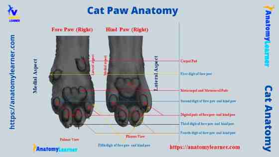
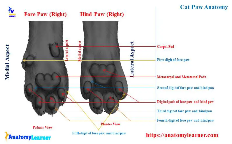
Cat paw anatomy diagram
Finally, I will show you all the structures from the cat paw anatomy with a labeled diagram. Here, I will focus on the external features and some of the internal features of the cats’ paws.
The labeled diagram shows the bones, some muscles, and some of the vessels from both the front and hind paws of the cats. You may find more paws labeled diagrams on the social media of anatomy learners.
Frequently asked questions on cat paw anatomy
Here, I will enlist the most frequently asked questions from the cat anatomy learners. I hope you will get the answers to your inquiries below.
What is the carpal pad for cats?
The carpal pad is the extra element in the structure of the forepaw of the cats. You will find this extra pad in each forepaw of a cat where the accessory carpal bone is present.
The structure of the carpal pad of the cat’s fore paw is similar with the other paws like metacarpal or digital pads. You will find a smooth and pigmented external surface in the carpal pads of a cat.
Why do cats have carpal pads?
As you know, there is an extra pad present in the forepaw of a cat. This is due to the accessory carpal bone in both the right and left foreleg. This accessory carpal bone covers by the digital cushion and forms the extra pad in a cat.
What is the extra pad on a cat paw?
The carpal pad is the extra pad on a cat’s forepaws. But, you will not find any extra pads in the cat’s hind paws.
What should cat paw pads look like?
What is the special about cats’ paws?
All the paws possess specific bones, muscles, blood vessels, and nerves. The external surface of the cats’ paws possesses some special features. It contains some pads – carpal, metacarpal, and digital pads. The most special feature of the cats’ paws is the presence of carpal pads in both right and left forepaws.
Do all cats have a carpal pad?
Yeah, all the cats have a carpal pad in their forepaw. This is the normal structure of the forepaws of a cat.
These are very little information about the anatomical features of the cat paws. If you want, you may learn more about the paws from the video offered by the anatomy learner.
Conclusion
So, there are 4 paws present all together in the cats’ legs. I hope you got the basic idea of the cat paw anatomy from this article. All the anatomical features of the bones, muscles, vessels, and nerves are equally important if you want to learn cat paws perfectly.
There is little difference between the cats’ forepaws and hind paw anatomy. There is a carpal pad present in the forepaw, whereas there is no carpal pad in the hind paw of the cats. Again, there are five metacarpals in the forepaw and usually four metatarsal bones in the hind paw.