The different breeds of dogs have a significant variation in the forms and size of their skull. Sometimes, you may find a long and narrow skull or find a broad and short skull in a different breed of dogs. This short article will show you the different bones from dog skull anatomy and their description.
I will also enlist the special osteological features from dog skull anatomy with a labeled diagram. Again, I will provide you with a bit of information on dog skull muscles anatomy, dog mandible anatomy, and more.
Don’t forget to check out the frequently asked questions section, as you may get answers to your common inquiries there.
Dog skull anatomy
Before going to the detailed description of the dog skull bones, I would like to provide a list of special osteological features. Let’s check out the following unique osteological characteristics from the dog skull anatomy.
- The skull is oval elongated in shape but may vary highly from breed to breed (dolichocephalic, brachycephalic, and mesaticephalic).
- There is a frontal ridge on the dog’s skull.
- The basioccipital bone joins with the bulla tympani in the dog skull.
- You will find a robust and highly curved zygomatic process in the dog skull.
- The interparietal bone fused with the occipital bone before birth in a dog.
- Parietal bone also takes part in the formation of the roof of the cranial cavity in a dog.
- You will find an incomplete orbital rim in the dog skull.
- You will also find a socket in the premaxilla bone for the incisor teeth.
- There is also a well-developed socket for the canine teeth in the premaxilla bone of the dog.
- The two haves of the dog mandible fuses incompletely.
- You will find other special features (masseteric fossa) on the dog mandible. This is a depression on the lateral aspect of the ramus of the dog mandible.
So, this is concise information about the dog skull bones. If you want to learn more about the osteological feature, please continue this article. You will get detailed information on every single bone from the dog skull.
Dog skull bone anatomy
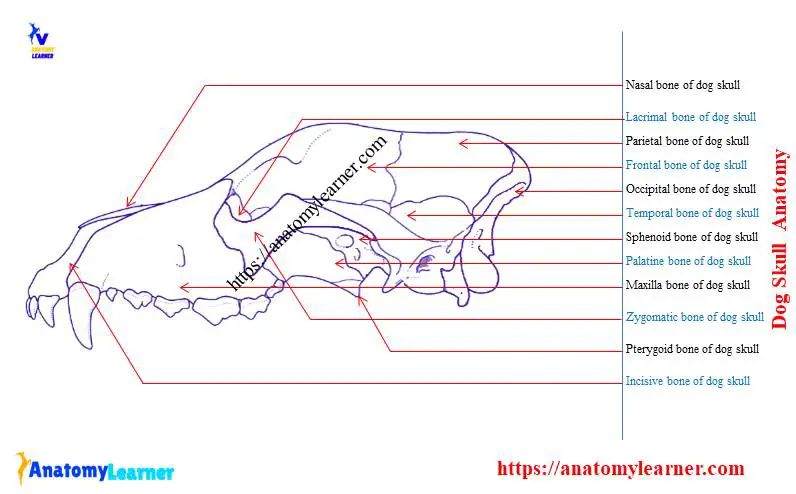
You might identify the following main bones from the skull of a dog. But, I will also show you the other different structures or processes of these main bones. So, what are these prominent bones of a dog skull?
Well, I will enlist the major bone from the dog skull. First, try to identify these bones and then go to the detailed description.
- An occipital bone of dog skull
- An interparietal bone of dog skull
- Parietal bones of a dog
- Frontal bones of a dog
- Temporal bones of a dog
- Different parts of sphenoid bones of dog skull
- Ethmoid bones of the dog
- Maxilla bone of a dog
- Incisive bones of a dog
- Palatine bones of a dog
- Pterygoid bones of a dog skull
- Nasal bones of a dog
- Zygomatic bones of dog
- Lacrimal bones of dog and
- Vomer bone of the dog skull
I hope the following dog skull anatomy diagram might help you to identify these main bones. So, want to know more about these dog’s bones?
The occipital bone of dog skull
The occipital bone is similar in position to that of the horse skull. There are two parts in the occipital bone of a dog – the basilar and the squamous part. The basilar part of the occipital bone is wide and joins with the tympanic bulla on either side. Again, the squamous part fuses with the interparietal bone before birth in a dog.
You will find an angular and prominent nuchal crest in a dog that directs caudally. There are two rough tubercles found just ventral to this nuchal crest. The ventral surface of these tubercles is convex from side to side and concave dorsoventrally.
You will find sizeable mastoid foramen on each side at the junction with the squamous part of the temporal bone. This foramen lies dorsal and rostral to the foramen magnum (large foramen of the dog skull).
There you will find flattened and widely separated condyles in dog skulls. You will also find a short condyloid canal at the medial side of each condyle.
There presence a very short jugular process in the dog skull compare to other animals. Near to the jugular canal, you will find a tiny canal for the hypoglossal nerve.
So, you might identify the following structures from the occipital bone of dog skull anatomy –
Basilar part of the occipital bone of a dog
Squamous part of the occipital bone of a dog skull
Prominent nuchal crest of dog
Tubercles for muscle attachement
Mastoid foramen of dog skull
Flattened condyles and condyloid canal
Jugular process and jugular canal of dog skull
Canal or foramen for the hypoglossal nerve
I hope you will identify all of these structures from the occipital bone of a dog skull. Let’s discuss the subsequent bone from the dog skull.
Interparietal and parietal bones of a dog
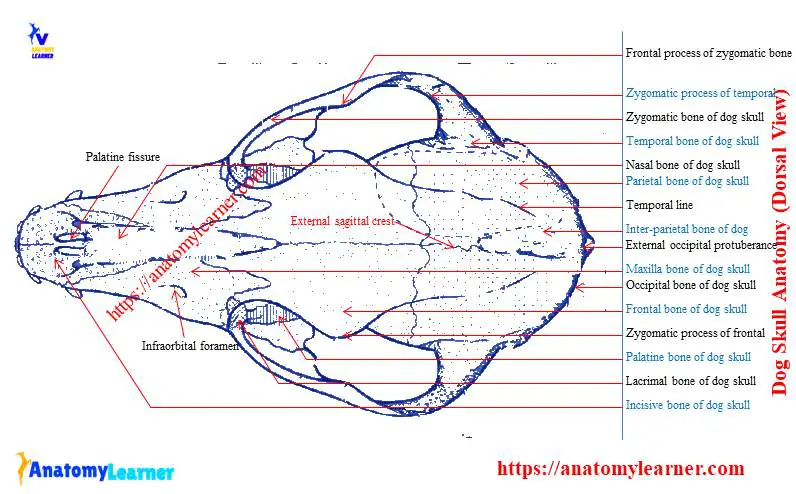
The interparietal bone of the dog skull fuses with the squamous part of the occipital bone before birth and forms the interparietal process. It contains the caudal part of the external sagittal crest.
The rostral end of the interparietal bone is narrower and thinner than the caudal end. It helps to form the central part of the osseous cerebellar tentorium internally.
The interparietal bone also takes part in the formation of the canal for the transverse sinus.
You will find a strongly curved and rhomboid-shaped parietal bone in the dog skull. This bone is very extensive and forms the more significant part of the roof of the cranial cavity of a dog.
You will find a prominent external sagittal crest at the junction of the right and left parietal bones in a dog. This external sagittal crest remains continue upon the frontal bone of the dog skull.
The ventral border of the parietal bone of the dog joins with the wing of the sphenoid bone. The external surface of this bone also takes part to form the temporal fossa. You will find some impression and ridges in the internal surface of the parietal bone of a dog.
So, what structures might you identify from these bones of a dog? You might identify the following parts from these bones –
External sagittal crest of the parietal bone of a dog
Area of interparietal and parietal bone of dog skull
The canal for the transverse sinus
Temporal fossa of dog
The external and internal part of the parietal bone of a dog
Please use the actual dog skull sample for identifying these structures from the interparietal and parietal bones of a dog.
Frontal bones from dog
In dog skull anatomy, you will find an irregular-shaped frontal bone that possesses some peculiar osteological features. You will find an orbital part, a temporal surface, a frontal squama, and a nasal part in the frontal bone.
The temporal line crosses the external surface of the frontal bone of a dog. It extends in a curve from the external sagittal crest to the zygomatic process.
The sagittal crest also separates the frontal squama from the temporal surface. The frontal squama of both sides together form a central depression and slope ventrally and rostrally.
You will find a very short zygomatic process in the dog skull. The supraorbital margin of the dog skull is incomplete, as seen in the pig skull. You may find supraorbital foramen on both sides of the supraorbital process of the dog skull.
The orbital and the temporal surface of the dog skull are very extensive. You will find two ethmoidal foramina in the frontal bone of the dog.
This bone articulates with the nasal bone and maxilla rostrally. Again, this bone also articulates with other bones like parietal, sphenoid, lacrimal, palatine, and ethmoid bones.
From this frontal bone area, you might identify the following structures –
Temporal line of dog skull
Short zygomatic process of dog skull
Supraorbital margin and supraorbital foramen of dog skull
Squama part of the frontal bone
I hope you will quickly identify these structures from the frontal bone of the dog skull. You may get help from the below-mentioned dog skull bone diagrams.
Temporal bones of a dog
The temporal bone of the dog forms a large part of the ventrolateral wall of the calvaria. There you will find petrosal, tympanic, and squamous parts in the temporal bone of a dog.
The petrosal part is also known as the pyramid or petrosum. It fuses around its periphery laterally to the medial surfaces of the tympanic and squamous parts. You will find the cerebellar fossa at the caudomedial aspect of the petrosal part of the temporal bone. Ventral to the cerebellar fossa, there is an internal acoustic meatus.
You will also find a short canal for passing the trigeminal nerve at the ventrorostral aspect of the internal acoustic meatus. The jugular foramen is located between the petrosal part of the temporal bone and the occipital bone of the dog. There is a distinct mastoid process at the petrosal part of the temporal bone.
The tympanic part of the temporal bone is the ventral portion and easily identified by its more significant component (bulla tympanic). At the rostral margin of this bulla you will find two large foramen – foramen lacerum and external carotid foramen.
The squamous part of the temporal bone contains a long, curved, zygomatic process. This process extends rostrolaterally, overlies the caudal half of the zygomatic bone, and forms the zygomatic arch. You will find a mandibular fossa at the ventral part of the base of the zygomatic process.
The inner temporal canal in between the squamous and petrous part forms passages for the temporal sinus. The squamous portion of the temporal bone of a dog overlaps the parietal bone and comprises a squamosal suture.
Now, I will show you the main identifying osteological features from the three parts of the temporal bones of a dog.
Sphenoid bone from dog skull
The sphenoid bones form the rostral two-thirds of the base of the cranial cavity in a dog. You will find two different parts (presphenoid and basisphenoid) in the sphenoid bone in the dog skull anatomy.
The more rostral bone with an orbital wing in a dog is a presphenoid, and the caudal bone with a larger wing is the basisphenoid bones in a dog. Now, I will discuss the essential osteological features of these two parts of the dog’s sphenoid bones.
The dorsal part of the body of the presphenoid bone is roofed over by the fusion of the right and left-wing that forms jugum sphenoidale. This jugum sphenoidale forms the orbitosphenoidal crest caudally. You will also find the rostral clinoid process at each side of the caudal end of the preshpenoid bone of the dog.
The body of the basisphenoid forms the base of the middle cranial fossa. It also helps to form the hypophyseal fossa. The wing of the basisphenoid bone is large, curves dorsally and laterally. At the base of this wing, you will find several foramina – oval foramina, foramen spinosum.
The pterygoid process is the ventrolateral extension of the basisphenoid bone in dogs. You will find the alar canal at the rostral part of the base of this pterygoid process.
The smaller opening of the alar canal is known as the caudal alar foramen, whereas; the larger one is the rostral alar foramen.
You will also find two pairs of the groove in the basisphenoid bone of the dog. The external pterygoid groove forms the small pterygoid canal.
The body of the basisphenoid bone articulate caudally with the basioccipital bone and forms sphenooccipital synchondrosis. Again, the basisphenoid bone articulates with presphenoid rostrally to form the intersphenoidal synchondrosis.
The ethmoid bone of the dog skull
The ethmoid bone is highly developed and located between the dog skull’s cranial and facial parts. You will find four different regions in the ethmoid bone of the dog.
A median perpendicular plates
Two ethmoid labyrinths covered by external laminae and
A cribriform plate
The perpendicular plate is the median vertical sheet of the bone that articulates with the vomer bone ventrally and septal process of the nasal and frontal bone dorsally. This perpendicular plate helps to form the bony nasal septum in a dog.
The cribriform plate is a deeply concave partition that articulates with the ethmoid notch of the frontal bone dorsally and the presphenoid ventrally and laterally. You will find several cribriform foramina on this plate that serve for the transmission of the olfactory nerves.
The cribriform plate forms the sphenoethmoid sutures with presphenoid bone ventrally. Again, it articulates with vomer to form the vomeroethmoid suture. Laterally and dorsally, the frontoethmoid suture is formed by the union of the cribriform plate with the medial surface of the frontal bone.
The ethmoid labyrinths form the bulk of the ethmoid bone in the dog skull. You will find the delicate bony scroll in the structure of both two ethmoid labyrinths.
Maxilla bone from dog skull anatomy
The maxilla is a very short but highly caudal bone in dog skull anatomy. You will find a body and four different processes in the maxilla bone of a dog.
The external surface of the maxilla bone of a dog has very prominent features, and that is the infraorbital foramen. You will find alveolar processes in the ventrolateral surface of the maxilla of the dog. There is a smooth elevation on the ventrolateral facial surface of the maxilla that is caused by the root of the teeth (alveolar juga).
The frontal process arches dorsally between the nasal bone and the orbit. The zygomatic process is largely hidden by the laterally lying zygomatic bone in the dog skull. Most of the hard plate of dog is formed by the transverse palatine process.
The dorsal surface of the palatine process forms the floor of the ventral nasal meatus. You will find the groove at the ventral surface of the palatine process, and it takes part in the formation of the roof of the oral cavity. The most caudal process of the maxilla is a small spur and is known as the alveolar process of the maxilla.
Again, the nasal surface of the maxilla is its medial surface and contains several crests. You will find the conchal ridge near the incisivomaxillary suture.
A palatine bone of dog skull
The palatine bone locates caudomedial to the maxilla bone in a dog. This palatine bone of the dog is divided into horizontal and perpendicular laminae.
The horizontal laminae or plate of the palatine bone is extensive and forms one-third of the hard plate. You will find a palatine surface, nasal surface, and a free concave caudal border in the horizontal lamina of the palatine bone.
There is a nasal crest at the nasal surface of the palatine bone that articulates with the vomer bone in a dog. You might identify the significant palatine foramen, minor palatine foramen, and palatine canal from the horizontal part of the palatine bone of the dog.
The perpendicular lamina of the palatine bone leaves the caudolateral border of the horizontal lamina. It helps to form the lateral wall of the nasopharyngeal meatus medially. Again, it forms the medial barrier pterygopalatine fossa laterally.
An incisive bone of dog skull
The incisive bone of a dog has a petite body and three processes. The body of the incisive bone compressed dorsoventrally. You will find the alveolar process, palatine process, and nasal process in the incisive bone.
The alveolar processes of a dog’s incisive bone have three alveoli for the three superior and inferior teeth. There are bony partitions between the alveoli known as the interalveolar septa.
You will find a curved and tapering nasal process at the dorsocaudal part of the incisive bone of a dog. The free rostral border of the nasal processes bound the bony nasal aperture. There are interincisive canals on the medial surface of each incisive bone of the dog.
The medial surface of the nasal processes articulates with the nasal bone to form the nasoincisive suture. Again, the incisive bone of the dog articulates caudally with the maxilla bone to form the incisivomaxillary suture.
The pterygoid bone of the dog skull
The pterygoid bone is a small, thin, slightly curved, and four-sided plate bone in dog skull anatomy. It articulates with the bodies of both presphenoid and basisphenoid bones of a dog.
You will find a pterygoid hamulus that extends from the caudoventral angle of the pterygoid bone. There is a smooth concave pterygoid fossa at the medial surface of the pterygoid bone of the dog.
The pterygoid bone forms a squamous suture with the pterygoid process of the sphenoid bone caudally. The name of this suture is pterygosphenoid suture.
Zygomatic bone from dog skull
The zygomatic bone forms the rostral half of the zygomatic arch in dog skull anatomy. It is a very long and strongly curved bone in a dog.
You will find two surfaces and two processes in the zygomatic bone of a dog.
The lateral surface of the zygomatic bone of a dog is convex longitudinally and transversely. There is a concave medial or orbital surface in the zygomatic bone of a dog.
The dorsal border of the zygomatic bone is convex and free rostrally. It takes part in forming the orbital margin of the dog.
The zygomatic bone articulates with the maxilla bone to form the zygomaticomaxillary suture. Again, at the rostral edge of the orbit, it forms the lacrimozygomatic suture by joining with the lacrimal bone.
Other bones from the dog skull
You might also know the detailed anatomical features of the vomer, lacrimal, and nasal bones from the dog skull. The lacrimal bone locates at the rostral margin of the orbit in a dog. This lacrimal bone of the dog is triangular in outline and pyramidal in shape.
You might identify the following osteological features from the lacrimal bone of a dog –
Frontal process of dog
Lacrimal canal of lacramial bone
Fossa for the lacrimal sac
The lacrimal bone of the dog articulates with the
frontal bone and forms frontolacrimal suture. Again, this bone rostrally articulates with the maxilla bone and forms a lacrimomaxillary suture.
The vomer of the dog skull anatomy is an unpaired bone and forms the caudoventral part of the nasal septum. This bone also contributes to forming the roof of the nasal concha of the dog.
The caudal end of the vomer bone is narrow and deeply notched. You will find the wings at the horizontal part of the vomer bone of a dog.
The nasal bone is long, slender, and narrow caudally in a dog. The external surface of the nasal bone varies in size and shape. A mucous membrane covers the ventral or internal surface of the dog’s nasal bone.
For more dog bone-labeled diagrams, you may follow anatomy learner on social media.
Mandible of the dog
The mandible of the dog possesses some peculiar features. I will provide the unique features of the dog mandible with a diagram.
The two halves of the dog mandible don’t fuse completely. You will find six alveoli for incisor teeth and two for the canine teeth in the body of the dog’s mandible.
The incisor alveoli increase in size from first to third. You will find the ventrally and caudally directed canine alveoli in the body of dog’s mandible.
The ventral border of the molar part is convex in its length and is thick and rounded. You will find very short interalveolar space in the body of dog’s mandible.
The rostral part of the mandible turns medially and posses two or three mental foramina. Do you know which one is the largest mental foramen in a dog? The middle one is the largest mental foramen in a dog that locates ventral to the septum between the first two cheek teeth.
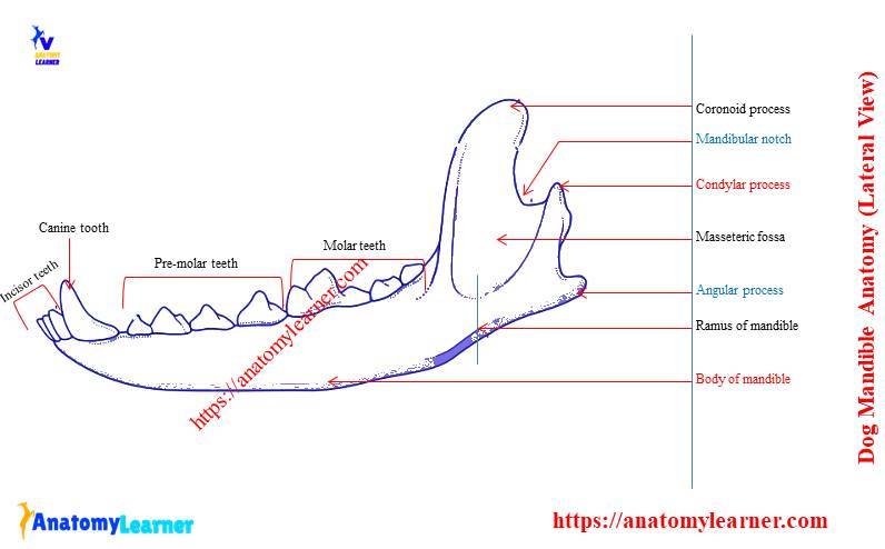
The ramus of the mandible is the caudal non-tooth-bearing verticle part of the bone. You might identify the following osteological features from the dog mandible.
Coronoid process of the dog mandible
Condyloid process of the dog mandible
Mandibular notch of dog mandible
Masseteric fossa of dog mandible
Angular process of the dog mandible
The angle of the dog mandible
The mandibular foramen and mandibular canal of dog
The ramus of the dog mandible is relatively small, and the lateral surface contains deep masseteric fossa. Again, the medial surface is convex and marked by the mandibular foramen. The caudal opening of the dog’s mandibular foramen is mandibular canal.
You will find a transversely elongated, sagittally convex articular process in dog ramus. The name of this process is the condyloid process. It forms the temporomandibular joint by articulating with the mandibular fossa of the squamous temporal bone.
Frequently asked questions on dog skull bone
So, in this part of this article, you will get the answers to the frequently asked question on the dog skull anatomy.
Is it normal for dogs to have a bump on their head?
Yeah, it is normal for some breed dogs to have a bump on their head. It is a bony prominence on the parietal and frontal bones of a dog skull.
What are the four skull types for dogs?
These dogs, which have a long but narrow skull, are designated as dolichocephalic. Other dogs have broad but short skull are termed as brachycephalic. And the intermediate form skull is the mesaticephalic skull.
How many skull bones do dogs have?
The number of bones in the skull of a dog varies with a different breed. But averagely, you may find forty bones in the skull of a dog.
What forms the apex of the skull in dogs?
What dog has the thickest skull?
Why does my dog have a ridge on his skull?
The interparietal bones fuse with the squamous part of the supraoccipital bone before birth. It forms the interparietal process that posses the external sagittal crest. This crest also continues with the subsequent bone (frontal) of the skull of a dog. So, you will find a ridge on your dog’s skull.
Do dogs have occipital bone?
Conclusion
I hope this simple guide might help you to learn the dog skull anatomy with labeled diagrams. I always suggest practicing all the osteological features of the dog skull bone with the actual sample along with the provided diagrams.
Don’t forget to learn the detailed anatomy of some major bones like occipital, interparietal, frontal, temporal, and sphenoid bones from the dog skull anatomy. For more updates on dog skull bones, you may stay connected with the anatomy learner.