There are considerable differences exist between mammals and chicken kidney anatomy. You will also find a significant variation in the formation and excretion of urine in chickens compared to mammals. Here, I will discuss the detailed anatomy of a chicken kidney with a labeled diagram.
You will also find a chicken urinary system organs diagram in this article. That might help you understand the unique features compared to a mammal. Finally, don’t forget to check the questions and answers sections on chicken kidney anatomy.
Nice, let’s get started to learn the anatomical facts of the kidney of a chicken or bird or any avian species.
Chicken kidney and urinary system
Before going to the detailed anatomy of a chicken kidney, I would like to provide some peculiar anatomical facts from the urinary system. These might help you get the basic idea of the urinary system organs of a chicken or bird or any other avian species.
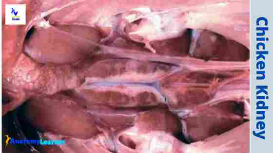
The kidney of a chicken is a brown-colored elongated structure that locates symmetrically on either side of the vertebral column below the pelvis and synsacrum.
There are fine lobulations present on the surface of the kidney and are usually divided into three major parts – cranial, middle, and caudal.
Each ureter of the chicken urinary system is divided into the renal part and the pelvic part. The renal part is embedded chiefly into the substances of the kidney. In addition, the pelvic portion of the chicken ureter runs from the caudal end of the kidney to the cloaca. Thus, both the ureters of a chicken opens at the dorsal aspect of the urodeum.
In chicken, you will not find any urinary bladder as in mammals. This feature reduces the bodyweight of a chicken and thus facilitates flight. The urinary component of chicken excreates together with feces.
Chicken kidney anatomy
There are right and left kidneys present in the chicken urinary system. I hope you might get interested to learn the chicken kidney anatomy with me. I will try to present the anatomical facts of a kidney quickly and concisely.
So, what I will cover here.
- The actual location of the kidney of birds or chickens,
- Forms of kidney of chickens (size, shape, and color), and
- Structure of chickens kidney
Make sure you have a good piece of knowledge on the skeleton of the chicken (especially about the synsacrum) and lung location.
Chicken kidney location
The two kidneys of a chicken symmetrically place on either side of the vertebral column. Therefore, they remain in contact dorsally with the pelvis and synsacrum.
As you know, there are deep bony crypts present in the ventral aspect of the pelvis and synsacrum of a chicken. The kidneys logs with these deep bony crypts of the chicken’s pelvis and synsacrum.
The caudal extremity of the chicken kidney reaches nearly to the caudal end of the synsacrum. Again, the cranial extremity extends just beyond the end of the synscarum and reaches the lung of a chicken.
In addition, the length and width of the kidney may vary within the chicken species. Averagely, the length is about seven centimeters, and the greatest transverse width is about two centimeters.
Color and shape of kidney – the standard color of a chicken’s kidney is brown. In addition, you will find an elongated rectangle kidney in the urinary system of chickens.
The shape and structure may vary in other different avian species (like in duck, pigeon) compared to chickens. You will find a little information on the kidney structure of a duck and pigeon at the end of this article.
Chicken kidney structure
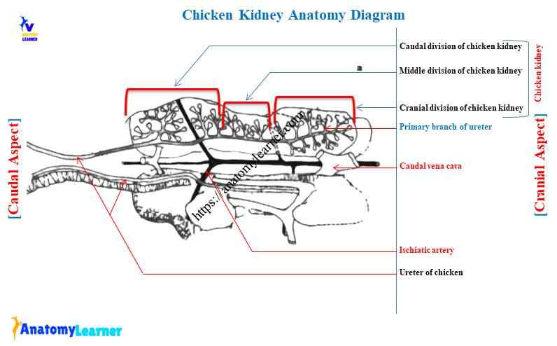
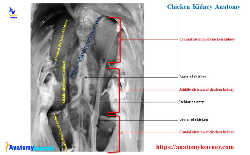
In the chicken kidney, you will find three major renal divisions that connect by a parenchymal bridge. Fine, let’s know what these renal divisions of chickens kidney are –
- Rounded cranial division,
- More slender cranial division, and
- The expanded and irregularly shaped caudal division.
The surface lobulation and the number of pyramids of a kidney may vary from animal to animal. For example, in chicken, the surface of each of the three large lobes of the kidney presents fine lobulation. So, the numerous small lobes club together and provide irregular appearances on the surface of the kidney.
You will find a groove on the dorsal surface of the boundary between the cranial and middle divisions of a kidney. This group housing the external iliac artery of a chicken. In addition, there is a groove on the ventral surface of the boundary between the middle and caudal divisions that are housing the ischiatic artery.
You will also find some other groove that forms by the following structures on the ventral surface of the kidney.
External iliac vein of chicken
Caudal renal vein of chicken,
Caudal portal vein and ureter of a chicken
“The ventral surface of the avian kidney is in contact with the paired abdominal air sacs and with the wall of the peritoneal intestinal cavity.”
The ischiatic and several spinal nerves lie between the renal divisions of a chicken’s kidney.
You will also find the small dome-shaped bulge renal lobule on the surface of the chicken kidney. So, let’s discuss the structure of renal lobes and lobules of a kidney from the avian urinary system.
Renal lobe and lobule of kidney
In the functional term, the chicken kidney anatomy divides into the renal lobes and renal lobules based on the branching pattern of the ureter. The renal lobes are the portion of the kidney medulla that drains into the second branch of the ureter. But, the renal lobe is not so distinct grossly in any avian species.
The lobule is the area of tissue that is wedged between the terminal branch of the renal portal vein. Some lobules project from the surface of a kidney as a small, round structure. But, most of the lobules remain deep to the surface of the kidney.
Each lobule of the bird kidney includes both medullary tissue and cortical tissue. The medullary component of each renal tubules consists of a cone-shaped bundle of medullary collecting tubules enclosing blood vessels and a loop of Henle of a juxtamedullary nephron.
An efferent vein (intralobular vein) of the renal portal system passes through the center of each lobule. In addition, there is an intralobular artery (afferent artery) that supplies the renal lobules of a chicken.
The interlobular artery gives afferent arteriole that each ramifies to form a rete mirabile (glomerulus). Post-glomerular efferent arteriole gives rise to capillaries that anastomose with the capillaries of the renal portal system. Again, these capillaries together forming the peritubular capillary network that surrounds the renal tubule.
Cortex and medulla of bird kidney
The cortex of chicken kidney anatomy forms by the broad region of the renal lobules. So, you will find cortical and medullary types of the nephron in the cortex of the bird kidney.
Again, the medulla forms by the medullary region of the renal lobules. Therefore, it consists of a conical bundle of collecting tubules assembled into the pyramid-like cone-shaped tufts of the ureter. It also includes a loop of Henle of medullary type of nephrons.
Structure of chicken nephron
You will find two types of nephrons in the chicken kidney anatomy. The majority have no loop of Henle and occurs in all regions of the cortex except near the medulla of the kidney.
These are the nephrons of cortical type and are reptilian in form.
The rest have a loop of Henle remain in the region of the cortex near to the medulla. These are the nephrons of the medullary type and are mammalian in form.
Renal corpuscle of avian kidney
The renal corpuscle is always present at the beginning of the chicken nephron. As in mammals, the renal corpuscle of a chicken consists of a glomerulus and thin-walled Bowman’s capsule.
The glomerulus is the capillary tuft that arises from the afferent glomerular arterioles. Again, the renal corpuscle of chicken is small but more numerous than those of mammals.
You will find two layers (visceral and parietal) in the Bowman’s capsule of a chicken. The visceral layer of this capsule lies against the capillary loop.
A single layer of the flattened epithelial is present in the parietal layer of a chicken Bowman’s capsule. In addition, you will find podocytes in the visceral layers of the capsule that possess the branching processes. It extends to the basal membrane and forms the part of the barrier responsible for glomerular ultrafiltration.
Type of nephrons in chicken kidney
The cortical type of nephrons is more outstanding in number in avian kidney anatomy. For example, you will find a small spherical renal corpuscle in the cortical nephron of a chicken. It joins the proximal convoluted tubule by a very short neck.
The proximal convoluted tubule forms about half of the total length of the chicken nephron. It makes several convolutions but essentially has three main limbs. The diameter of these proximal convoluted tubules is similar to the renal corpuscle.
Again, the distal convoluted tubule of chicken nephron forms the compact convolution near the central interlobar vein. A short connecting tubule also presents in the cortical type of nephron of a chicken. But you will not find any distinct loop of Henle in the cortical type of nephron of a chicken.
On the other hand, the medullary nephron of a chicken has more substantial renal corpuscles and a well-defined loop of Henle. The renal corpuscle of the medullary type nephron is more significant than that of the cortical type of nephron.
The proximal convoluted tubule is the same as in the cortical nephron of a chicken. Again, the intermediate segment forms a loop of Henle that dips into the medulla. The distal convoluted tubules form a few compact colis near the central vein.
Tubules and collecting ducts of the kidney
You will find some differences in the tubule structure of chicken kidneys compared to mammals. The length and the diameter of the loop of Henle are less in a chicken compare to the mammal.
“You may skip this part of the article as you already got an idea on the tubules and collecting duct of different types of chicken nephrons.”
The convoluted tubule of the cortical nephron consists of a proximal tubule, a short and variable intermediate segment, and a distal tubule. Thus, a clearly defined loop of Henle is lacking in the cortical nephron of a chicken. The proximal tubules of cortical nephron also lack cuboidal epithelial lining.
In contrast, the medullary nephron consists of a proximal convoluted tubule, a proximal straight tubule, a thin tubule, a straight distal tubule, and a convoluted distal tubule. Finally, Henle’s descending loop comprises a straight proximal tubule, thin tubule, and the initial segment of the straight distal tubule.
The distal convoluted tubule of chicken nephron opens into the peripherally located collecting tubules. At the tip of the medullary cone, the medullary connecting tubules from several renal lobules.
The juxtaglomerular apparatus of the chicken nephron
As in mammals, the juxtaglomerular apparatus of chicken locates at the vascular pole of the renal corpuscle. You will find the following components in the chicken juxtaglomerular apparatus –
- Macula densa of chicken juxtaglomerular apparatus,
- Epitheloid or juxtaglomerular cells, and
- Extraglomerular mesangial cells or goormaghtigh cells.
The cells of macula densa of chicken juxtaglomerular apparatus are chemosensitive, monitoring the sodium concentration in the blood in the different arteriole.
Urine formation in chicken
Due to the shorter and thinner structure of the loop of Henle, chicken has a relatively limited capacity for concentrating urine. The uric acid forms as an end product of purine metabolism in chicken. You will find mucin and mucopolysaccharide containing materials that excrete from the epithelium of the ureter block of a chicken.
These materials aggregates and precipitate as a large urate crystal. The ureter ultimately deposits a dense, mucus stringy urine into the urodeum of the cloaca.
“Chicken excretes their nitrogenous waste as uric acid, whereas mammals excrete it in the form of urea.”
There is no facility to store urine as there is no urinary bladder in the chicken. In chicken, urine and feces expel together. The urine component consists of a liquid portion and a white paste containing concentrated uric acid.
Frequently asked questions on the chicken kidney.
You may get your desire questions and answers on chicken kidney anatomy here in this part of the article. I will try to cover all the questions asked by the chicken anatomy learners.
Does a chicken have a kidney?
Yes, a chicken has a kidney that possesses a considerable variation in its structures compared to mammals. You will find lobulation finely on the surface of both the right and left kidneys of chickens.
Each kidney divides into three divisions roughly equal length – the cranial, middle, and caudal division. In addition, you will find different grooves on the dorsal and ventral surface of the chicken’s kidney. These grooves contain different arteries, veins, and ureters of the chicken.
If you want to learn the details of the anatomy of the chicken’s kidneys, I suggest you read the whole article.
How many kidneys does chicken have?
You will find two large and well-defined (right and left) kidneys in a chicken urinary system. The right and left kidneys possess three different divisions – the round cranial division, slender middle division, and the expanded caudal division.
The anatomical structures of both the right and left kidneys are similar in chickens. Both kidneys possess the cortical and medullary types of nephrons.
You might learn the detailed structures of the nephron of a chicken to compare it with mammals.
What is the function of the kidney in the chicken?
The avian kidney has a defined function to produce, convey and excretes urine directly to the urodeum of the cloaca. Again, the renal corpuscle of the chicken nephron designs for the glomerular ultrafiltration.
Where are kidneys located in chicken?
The chicken kidneys locate on either side of the vertebral column in contact dorsally with the pelvis and synsacrum. You will find the cranial extremity of the chicken’s kidney just beyond the synsacrum, and it reaches the lung.
In addition, the caudal extremity extends nearly to the caudal end of the synsacrum bone of the chicken skeleton. You will find contact with the abdominal air sacs on the ventral surface of each kidney.
What is the shape of the kidney of the chicken?
The shape of the chicken’s kidney is an elongated rectangle, and the surface posses some fine lobulation. But the form of the kidney may vary within the other avian species.
Does the bird have lobulated kidneys?
Yes, a bird has lobulated kidneys, but the lobulation is not as distinct as mammals. Instead, they have lobulation finely on the surface of each right and left kidney. This is probably due to the fusion of the numerous kidney units.
In chicken, the numerous small lobules join together to form a lobe and give an irregular appearance on the surface.
How to differentiate duck and pigeon kidneys from chicken?
In duck, the kidneys are relatively long at craniocaudal and narrow transversely. The transverse width tapers progressively towards the cranial end of the duck kidney.
You will find a narrow zone in the cranial division of the duck kidney and a great length in the caudal division.
Again, the pigeon kidney has an expanded cranial division, slender middle division, and a minor caudal division.
Does ostrich have a urinary bladder?
No, there is no urinary bladder in ostrich. But you will find a dilated pouch in the ureter of an ostrich responsible for storing the urine until discharged.
Blood supply to chickens kidney
Three pairs of renal arteries supply blood to the chicken kidney. The three pairs of renal arteries of a chicken are –
Cranial pairs – arise from the aorta,
The middle pairs arteries – arises from the ischiatic artery and extends cranially through the middle division,
The caudal pairs arteries –arises from the ischiatic artery of a chicken.
Chicken urinary system organs diagrams
Again, I will show you all the organs from the chicken urinary system with a labeled diagram. Here, I tried to show you the right and left kidneys, ureter, arteries, veins, and other parts of the chicken urinary system.
You will get more updated labeled diagrams on chicken or avian urinary system here on social media.
You may also read the following article from anatomylearner.com (chicken or bird anatomy related articles)
- Birdwing anatomy with labeled diagram
- Anatomy of the digestive system organs of a chicken
- A chicken respiratory system with actual samples and diagrams
You will get more articles on the chicken or other avian species on the avian anatomy section of the anatomy learner.
Conclusion
Considerable differences exist between mammals and chicken kidney anatomy. As a veterinary anatomist, you might know the exact location and structure of chicken kidneys and others. This article might help you to learn the location and structure of the chicken kidney. In almost all avian species, you will find the same anatomical facts of the kidney.
Now, you should make a difference between the anatomical facts of chickens and mammalian kidneys. Make sure you also know the other interesting anatomical facts of others organs from the chicken urinary system.