
Bone is a special connective tissue that contains cells, fibres, ground substances and inorganic bone salts. In bone, you will find two types of substances – compact and spongy substances. In this short article, I am going to describe compact bone histology with a labelled diagram.
Hello dear anatomy lover, do you want to learn the details histology of compact bone? You are in the right place now, and I will help you learn bone histology with real slide pictures. Here, you will find the most important identifying points for compact bone histology slide under the light microscope.
After reading this short article, you will differentiate the compact bone histology slide from the spongy bone histology slide. Again, I will provide a compact bone histology drawing for you at the end of this article.
So, do you interest to learn bone histology with me? Let’s continue this article and know the compact bone structure.
Compact bone histology
From the compact bone histology slide, I will enlist some important histological features that you might identify at the laboratory. First, you should find out these features or structures from the bone slide pictures.
- #1. Internal circumferential lamellae of compact bone
- #2. External circumferential lamellae of compact bone
- #3. Overall osteon or Haversian system of bone
- #4. Center canal or Haversian canal of an osteon
- #5. Concentric lamellae of osteon or haversian system
- #6. Lacunae in concentric lamellae of haversian system
- #7. Canaliculi in the lacunae of osteon
- #8. Cement line in osteon structure
- #9. Interstitial lamellae in between the Haversian system or osteon structure
Fine, try to identify these structure from the bone slide labelled image.
Identification points of the compact bone slide
Do you want to identify the compact bone slide under the light microscope? Okay, I will provide you with the essential identification points for the compact bone histology slide under the light microscope.
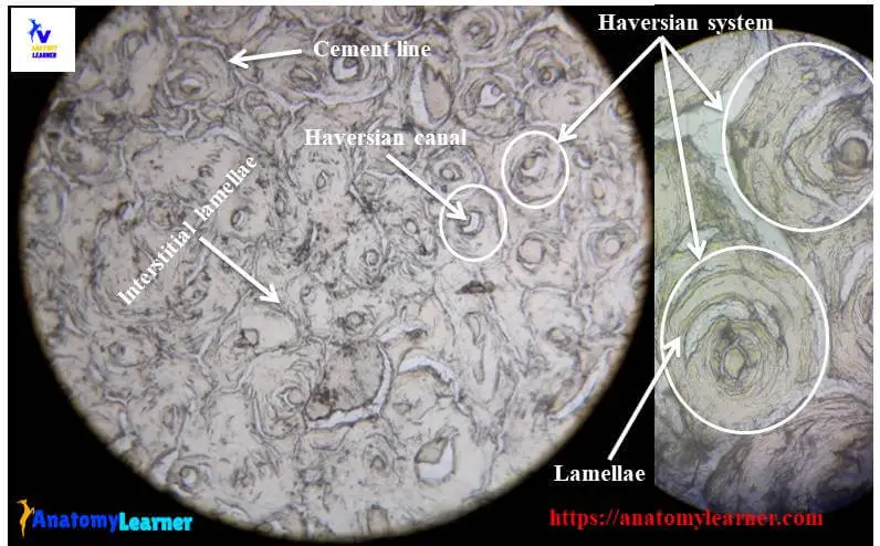
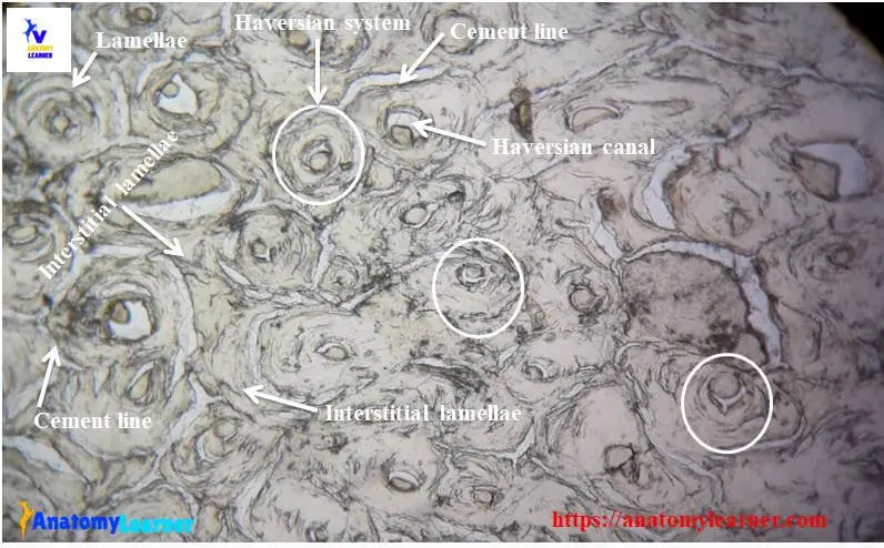
#1. The sample section of the tissue shows numerous ring-like structures (known as osteon or Haversian system of bone structure)
#2. Presence of Haversian or central canal at the centre of each osteon structure.
#3. There are concentric lamellae arranged around the central canal of the Haversian system.
#4. Presence of elliptical-shaped structure (known as lacunae) which contain the bone cells – osteocytes
#5. Some canaliculi radiate from the lacunae and hold the cytoplasmic process of the osteocyte.
#6. Presence of cement line around the osteon structure.
#7. There are interstitial lamellae in between the Haversian system of the sample tissue structure
So, this is a slide of compact bone histology.
Compact bone histology slide structure with diagram
Do you want to learn the details of the histology of compact bone with labelled diagram and authentic slide images? Good, here in this part, I am going to describe the structure of compact bone. In compact bone, you will find the three bone lamellar system in an orderly manner –
- #1. Circumferential system
- #2. Haversian system and
- #3. Interstitial system of compact bone
Circumferential system of bone structure
The outer circumferential system consists of circular lamellae of the bony matrix that lie immediately beneath the periosteum. Again, in the inner circumferential system, you will find the same circular lamellae of the bony matrix adjacent to the endosteum.
More lamellae are found in the outer circumferential system, but fewer numbers are located in the inner circumferential system of bone structure. You will also find the Haversian and interstitial system in between the outer and inner circumferential system.
Haversian system or osteon
This (Haversian system or osteon) is the structural unit of a compact bone matrix. They are the long cylindrical and branching structural unit that lies parallel to the long axis of the bone shaft.
Each of the osteon or Haversian systems contains a centre canal or Haversian canal at the system’s centre. This Haversian canal surrounds by the different layers of concentric lamellae, which are the thin plate of bone.
The Haversian canal is lined by the endosteum and contains blood vessels, lymphatics, nerves and connective tissue. The diameter of the Haversian canal in different osteon may vary. The Haversian canals communicate with each other through a transverse channel known as the Volkmann canal (not shown in the diagram).
In the concentric lamellae, you will find some almond or elliptical structure or space that contains osteocytes. These structures or spaces are known as the lacunae of the Haversian structure.
You will also find some tiny canal or canaliculi radiate from the lacunae in a different direction that anastomoses with the canaliculi from other lacunae. These canaliculi also contain the cytoplasmic processes of osteocytes.
Some of the canaliculi directly open into the haversian canal of the haversian system and the marrow cavity of the bone. You will find another structure in the Haversian system – the cement line. The cement line is the refractive line of the bony matrix around the Haversian system.
Interstitial system of compact bone
You will find some small irregular area of bone in between the Haversian system. There you will find some lamellae (known as interstitial lamellae). In these lamellae, you will find the same histological features you saw in Haversian system’s lamellae.
Compact bone histology drawing
Do you want to draw the compact bone histology slide picture? Well, follow the slide picture and try to draw the compact bone structure picture. Here, I am going to share a compact bone histology drawing with you.
There may find some mistake, if you found any error then let me inform. If you need more real slide pictures of the compact bone slide, then you may follow anatomy learner on social media.
Spongy or cancellous bone histology
If you want to learn spongy or cancellous bone histology, then you may read the article from the anatomy learner article page. You might read the other different article related to veterinary anatomy and histology from anatomy learner –
#1. Histology of oesophagus with slide pictures
#2. Histology of kidney with slide pictures
Conclusion
Hope you got the best guideline to learn compact bone histology with real slide pictures and labelled diagram. If you think this is the best guide, you may share this article with your friends who want to learn bone histology with slide images and labelled diagrams.
Let’s follow anatomy learner on social media and get more updates on the articles and images.