
In the last article, I described the compact bone histology with labeled diagram and real slide pictures. This short article will describe the spongy bone histology and labeled diagram and real slide pictures.
Hey there, welcome back again to anatomy learner and thank you so much for getting into this article. If you continue to learn the bone histology description, then this article is for you. Here in this article, you will find a short but essential description of spongy bone with slide images. You will also find the spongy bone histology slide drawing tutorial at the end of this article.
So, if you are interested to learn spongy bone histology a then continue this article till the end.
Spongy bone histology
You already know that bone composed of two substances, and they are – compact substance and spongy substance. From the spongy bone histology slide, we will identify the following important histological features under the light compound microscope.
- #1. Connective tissue with blood vessels surrounds the periosteum of spongy bone
- #2. Periosteum of the spongy bone sample
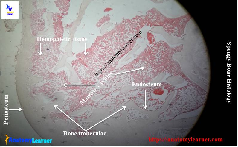
- #3. Bony trabeculae in spongy bone structure
- #4. Lacunae and osteoblast in the lacunae (at bony trabeculae)
- #5. Marrow cavities in spongy substance of bone
- #6. Blood vessels and hemopoietic tissue in marrow cavities
- #7. Compact bone (if present in the provided sample)
Okay, let’s find these histological features from the spongy substance from the provided slide pictures and the diagram.
Identification points of spongy bone slide
Do you want to identify the spongy bone histology slide under the light compound microscope? Fine, I will help you to identify the spongy bone slide’s features. I hope the following identification points might help you to identify the spongy substance of bone from the slide.
#1. The sample tissue shows numerous bony trabeculae
#2. Some of these trabeculae are single, and some are branched and anastomose with other branches of trabeculae
#3. Presence of marrow space that surrounds with bony trabeculae and contains bone marrow
#4. There are numerous blood vessels and hemopoietic tissue at marrow space (replace 3)
#5. Osteocytes are embedded in the bony matrix of the trabeculae in the sample tissue
#6. Presence of osteoblast and osteoclast on the surface of the bony trabeculae
#7. There are no Haversian systems in the sample tissue section
So, this is a spongy bone histology slide.
Histological description of spongy bone
So, do you want to know the details on spongy bone histology with slide pictures? Great, let’s continue this article to learn more about spongy substance of a bone structure.
Spongy bone aslo known as the cancellous bone (please keep in mind). This spongy bone is made of slender bony trabeculae that may be single or branched. The branched bony trabeculae anastomose with other branches of trabeculae. These trabeculae enclose the irregular marrow cavities between them, which contain blood vessels.
Each trabecula of the spongy bone is made of several parallel lamellae of the bony matrix. These lamellae of the bony matrix consist of lacunae which contain osteocytes.
You will find the marrow space between the trabeculae in the spongy bone structure. These marrow spaces contain blood vessels and hemopoietic tissue. You know these hemopoietic tissues are responsible for producing the new blood cells.
The bony trabeculae are covered by the vascular endosteum externally. Here in the trabeculae, you will also find the osteoprogenitor cells, osteoblast and osteoclast cells. You know, the osteoprogenitor cells are responsible for giving rise the osteoblasts.
Other histological features of spongy substance
You will find the periosteum that surrounds the trabeculae of spongy bone externally in the spongy histology slide. This periosteum merges with adjacent dense irregular connective tissue with numerous blood vessels. Again, inferior to the periosteum, the bony trabeculae may join with a thin layer of compact bone in a different section. These sections may contain the primitive and the mature osteon or Haversian systems with concentric lamellae.
Spongy bone histology drawing
Fine, it is not such a hard job to draw spongy bone histology slide images. You might draw what you have seen under the light microscope. Here, I am going to share the simple drawing of a spongy histology slide image with you. You may follow this guide but try to draw a better spongy structure then this drawing.
If you need more histology drawing tutorial and more real slide images, then you may follow anatomy learner on social media. You will get more updates on different slide’s photos there from anatomy learner.
Compact bone histology slide
If you want to learn about compact bone histology, then you may read this article. This article will know the circumferential system, Haversian system, and Interistial system of compact bone histology in details. You will also find the essential identification points of compact bone slide under the light compound microscope. So, if you missed it, then go now and read this article.
Bone histology description
If you asked to answer the bone histology, you might write both compact substance and spongy substance histology. Again, you may ask only the Haversian system or compact bone features or spongy bone structure.
I think you have a great idea of the different bone cells – osteocytes, osteoblast, and osteoclast. If you want to know details on these bone cells, then you may read the article from anatomy learner. You will find the details guide to learn the osteoblast, osteocytes and osteoclasts with a labeled diagram.
Again, you might learn the bone formation processes (known as osteogenesis) in details. You know there are two processes of bone formation – intramembranous ossification and endochondral or intracartilaginous ossification. Again, you will also find the details guide of ossification here in the anatomy learner blog with step by step process images.
You might also read the other different article related to histology or anatomy from the anatomy learner blog –
#1. Details histology of different types of cartilages – hyaline, elastic and fibrocartilage with real slide images
#2. Histological features of loose connective tissue
#3. Identification points of dense regular and irregular connective tissue under a light compound microscope and their description
Conclusion
I hope you learn the basic histology of spongy bone with real slide pictures. If you think this article may help your friend who wants to learn bone histology, then share it.
Are these identification points of spongy bone histology slide enough for identification? If you find any mistake on spongy bone histology labeled slide pictures, then let me know. Finally, don’t forget to share this article and follow anatomy leaner for regular updates.