Cows are essential livestock that provides excellent value to their owner. The cow anatomy deals with the forms and structure of their particular organ. It is not possible to describe all the anatomical features of a cow in a single article. But, I will try to summarize all the external and internal anatomical features of a cow here in this article.
Hi, if you are a veterinary student, you might know a cow’s basic anatomy. Again, if you are a farm owner, you might also know the external body parts of a cow and their internal organs in a little to fine-tune your knowledge.
Here in this article, first, I will show you the different body parts of a cow with a labeled diagram. Then I will discuss the anatomy of some vital internal organs of a cow.
You should know the details of bones, muscles, digestive organs like stomach, respiratory organs, nerves, and veins from a cow. I will try to show you all with short information along with labeled diagrams.
Cow anatomy
I hope you know the definition of the term animal anatomy. This means the branch of biological science that deals with the forms and structure of the particular organ of domesticated animals. So, if you want to learn the cow anatomy, you might know the shape, size, location, composition of each particular organ of their body.
I hope you could understand the depth of the anatomical features of a cow. If you want to learn the details anatomical facts of a cow, I will suggest you learn system-wise. But, if you want to get a basic idea of a cow external and internal organs, you may continue this article.
It might be best if you covered the anatomical facts of the following different organs system of a cow –
A cow’s skeletal system deals with the description of bones (both the axial and appendicular skeletons).
Syndesmology of cow – deals with the description of joints from cow’ body.
Myology of a cow – here, you might learn the list of clinically essential muscles with their origin, insertion, and fiber direction.
The organ from the digestive system – in this system, you might learn the anatomical facts of digestive organs of a cow-like – tongue, esophagus, stomach, intestine, liver, pancreas, and more.
Respiratory systems organs – this system deals with the description of the lung and trachea of a cow.
Here, in the cardiovascular system of a cow, you might learn the location, description of the heart, and clinically essential vessels of a cow.
The nervous system of a cow – here, you will learn different parts of cow’ brain, cranial and peripheral nerves, courses of some vital peripheral nerves like – radial, median, ulnar, ischiatic, tibia, and fibula.
Other organs systems of cow – you will also learn the anatomical facts of organs from other systems of a cow (showing in diagrams).
External body parts of a cow
Okay, you should become familiar with the different external body parts of cow anatomy. This is very simple but important to know the terminologies of cow body parts. It is essential to judge the utility of a cow and also for the classification of the breed.
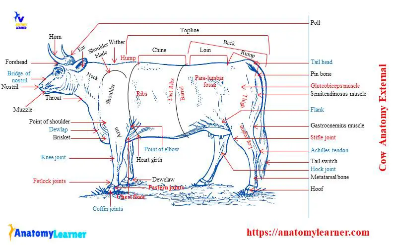
You might identify the different external body parts of a cow from the following five major regions. I will try to show you all of the body parts of a cow in labeled diagrams.
The external body parts from the head region of a cow – in this head region, you might identify the mouth, lip, cheek, chin, muzzle, forehead, poll, ear, eye, nostril, and other.
Different parts from the neck region of a cow – here, you will find the neck crest, dewlap, brisket, and jugular groove.
The external parts from the body or barrel of a cow – in this region, you will find many external features. The most important features are – hump, wither, loin, rump, pin bone, heart girth, flank, and others.
Different parts from the forelimb of a cow – you might identify the shoulder, point of shoulder, arm, forearm, knee joint, cannon bone, fetlock, pastern, and coffin joints.
The external features of the hindlimb of a cow – here in this region, you will find a point of the hipbone, thigh bone, leg bone, stifle joint, hock joint, Achilles tendon, and more.
If you dont know the terminologies of the external body parts of a cow and want to know more, you may continue the following part of the article. However, I have already described some of these terms previously in an external goat anatomy article.
Little on terminologies of cow body
You know, a cow’s mouth consists of lips, teeth, tongue, jaw, and dental pad. There is a well-developed cornual process present in a cow. This process may be back at a young age and yellowish black at an older age. But in some breeds of cow, you may find the pinkish-yellow cornual process.
There presence several rings on the tips or base of the horn. You may determine the age of a cow with the help of several rings on its cornual process.
You will also find the head crest in between the base of the cornual-process of a cow. Below the head crest and above the level of two eyes of a cow, there is a flat forehead. The poll of a cow is a bulging portion that lies at the top middle center of the forehead.
The chin of a cow lies below the lower lip, and it is fleshy and pinkish. Again, the muzzle is the projecting part of the head, including the mouth, nose, and jaw of a cow.
You will find a neck crest that lies between the head crest and hump. In addition, the hump is the bulging fleshy portion between the neck crest and back.
There is a well-developed dewlap in a tropical breed of cow. But you will find less developed dewlap in European cows.
There is a fleshy bulging brisket present between and in front of the forelimb of a cow.
You will find the wither of the cow just behind the hump and in between two shoulders. The back of a cow is the part between wither and the last rib.
You will find a well-developed flank region in a cow. This is a part of thick skin that hanging from the hindlimb and abdomen.
Internal body parts of a cow
In the internal body parts of cow anatomy, you will learn a lot of organs or structures. Here, I will discuss the most important organs with their identifying anatomical features.
But, you should know the detailed anatomical features of cow’s bones first. I will try to provide a basic idea of the different bones of a cow.
Great, let’s see the cow internal anatomy organs labeled diagram. I hope it will give you the basic concept of cow’s internal organs.
Cow anatomy bones
Unlike the other mammals, you will find the axial skeleton and appendicular skeleton in a cow. Here in this part of the article, you will get the anatomy of bones from a cow skeleton.
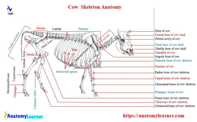
Bones of a cow from both axial and appendicular skeletons –
The bones of the forelimb of a cow – scapula, humerus, radius-ulna, carpal, metacarpal, and phalanges.
Hindlimb bones of a cow – include ilium, ischium, pubis, femur, tibia-fibula, tarsal, metatarsal, and phalanges.
The bones from an axial skeleton of a cow – include bones of the skull, vertebrae, ribs, and sternum.
Fine, let’s discuss the bones mentioned above from a cow skeleton in a little.
Bones of thoracic limb of a cow
The thoracic limb consists of four chief segments – thoracic girdle, arm, forearm (radius and ulna), and maneus. I will show you the anatomy of these thoracic limb bones of a cow.
The thoracic girdle of cow anatomy consists of a large, well-developed scapula and a small fused coracoid process. You will find a more regular triangular scapula in a cow than that of a horse. The scapula is relatively more expansive at the dorsal end and narrower at the ventral end.
There is a more prominent spine on the lateral surface of the cow’s scapula. This spine becomes less prominent at the ventral end and forms an acromion process. The coracoid process is short and rounded in a cow.
You will find a shallow musculosprial groove for the brachialis muscles in the humerus of a cow. The deltoid tuberosity of cow is less prominent than in horses. You will find a large greater tubercle that forms the point of the shoulder in a cow.
The radius of a cow is relatively short but broad than those of a horse. There is a marked increase in with and thickness distally. Again, the ulan of a cow is much less reduce bone than in the horse. You will find a complete, three-sided, strongly curved body in the ulna bone of a cow. The olecranon process is extensive and bears a more prominent tubercle.
The maneus of a cow consists of six carpal bones, a fused metacarpal, and four digits ( two digits are fully developed).
The radial and intermediate carpals resemble, in general, those of the horse. The body of the metacarpal bone of a cow is shorter than in a horse and relatively broader and flatter.
Nice, If you want to know more about the thoracic limb bone, you may read these articles.
Bones from the hindlimb of a cow
The pelvic girdle of the cow consists of the ossa coxurum and the sacrum bone. This oss coxae is the most prominent flat bone in a cow that consists of ilium, ischium, and pubis bones. These three bones meet to form a large acetabulum cavity that articulates with the head of the femur.
The ilium bone is almost parallel and forms a slight angle with the horizontal plane than in horse. You will find a main gluteal line in the cow ilium bone anatomy. Again, the coxal tuber is relatively large and prominent than those of a horse.
The acetabulum of the cow is smaller than in the horse. You will find two notches at the rounded rim of the cow’s acetabulum. In addition, the obturator foramen of the cow is large and elliptical with a thin and sharp medial border.
There is a relatively small and cylindrical body present in the femur of a cow. The lesser trochanter has a rough tuberosity that lies higher than that of a horse. You will find a small head in the femur of a cow than those of a horse.
There is a long, narrow, and very thick patella in a cow. The tibia resembles that of the horse rather closely but is somewhat shorter. You will find a less muscular line at the caudal part of the cow’s tibia bone.
The fibula of a cow consists of two extremities – proximal and distal. Unlike the other animals, the pes of cow consists of tarsal bones, metatarsal bone, and phalanges. You will find five pieces of tarsal bones in a cow. The calcaneus is longer and more slender than in the horse.
The dorsal longitudinal groove of the metatarsal bone is more profound. You will find a variable number of grooves at the plantar surface of the cow’s metatarsal.
Cow skull anatomy
The anatomy of a cow skull is somewhat different than those of a horse or other mammals. You will find a more pyramidal, shorter, but relatively broader skull in a cow.
You will find a quadrangular and more oversized cranium in cow’s skull. This is due to the presence of large frontal sinuses in a cow.
The frontal surface of the cow’s skull consists of frontal, nasal, and incisive bones. At the rostral part of the frontal surface, you will find a central depression on either side that posses a supraorbital groove and foramina.
There is a concise nasal part in the cow skull. The incisive bones of a cow are thin and relatively weak than those of a horse.
The lateral surface of the cow’s skull is more triangular than the horse that posses a temporal fossa. You will find a short, flattened, and weak zygomatic process at the lateral surface of the skull.
The orbital margin is completed by the frontal process of zygomatic bone in a cow. But in the canine skull, it is incomplete (exceptional features in dog). The pterygopalatine fossa is much larger, deeper, and distinct in the skull of a cow.
You will find double infraorbital foramen on the dorsal surface of cow’s skull at the level of the first cheek tooth. There is a facial tuberosity and curved line present in the skull.
There is a short and wide basal surface in the skull of a cow. In addition, the nuchal surface is extensive and somewhat pentagonal in outline. The occipital condyle remains limits cranially by the transverse ridge in a cow. There is a sizeable tympanic bulla present in the cow’s skull.
You may know the other different osteological features of a cow skull in detail here.
Vertebrae of a cow
You will find seven cervicals, thirteen thoracic, six lumbar, five sacral, and eighteen to twenty caudal vertebrae in the vertebral formula of cow anatomy. Now, I will show you the essential features of these vertebrae in a little. However, I have a detailed article on ruminant vertebrae here on anatomy learners.
The cervical vertebrae of a cow are much shorter than that of a horse. The articular processes are smaller than in the horse, and a plate of bone connects each two of the same side.
You will find a well-developed spinous process in the cervical vertebrae that is directed dorsally and cranially. There is a double transverse process in the third, fourth, and fifth cervical vertebrae of a cow. But the seventh cervical vertebrae possess a single, thick, and short transverse process.
You will find a thick ventral arch, less curved wing in the atlas of a cow. The axis is very short, and the lateral vertebral foramen is almost circular. The thoracic vertebrae of a cow are more prominent than those in a horse. You will find a longer body and a distinct constriction at the middle part of the thoracic vertebrae of a cow. The cow’s transverse process of the thoracic vertebrae is thick and strong, and possesses a rounded mamillary process.
There is also a constriction present at the middle of the lumbar vertebrae of a cow. These vertebrae are much larger than that of a horse and possess a rudimentary ventral crest.
You will find five segments in the sacrum of a cow. The spinous processes of these segments fuse entirely and form the median sacral crest in a cow.
There are well-developed caudal vertebrae in a cow than that in a horse. These vertebrae possess a groove for the median artery ventrally.
Ribs and sternum anatomy of a cow
There usually are thirteen pairs of ribs present in the cow skeleton anatomy. Out of these thirteen pairs, eight pairs are true ribs, and the rest are asternal ribs. They are generally longer, flatter, less curved, and less regular in form than in the horse.
In addition, the eighth, ninth, and tenth ribs are the longest and most comprehensive in cow skeleton. At the proximal end of the cow ‘s rib, you will find the exact structure of other mammals. You will find a coastal grove at the caudal border of the cow’s ribs.
The neck of the ribs is longer than those of a horse. Again, the articular surface of the tubercle is concave transversely, except on the last two or three ribs.
There are seven segments or sternebrae present in the sternum of a cow. The sternum of the cow is relatively wider, flatter, and longer than in the horse.
You will find a wedge-shaped manubrium in the cow’s rib that is compressed laterally. The body of the ribs widens craniocaudally. You will find several notches at the lateral border of the ribs for passages of the vessels.
Cow muscle anatomy
It is also essential to know the essential muscles anatomy of a cow for field practices. I will show you the different clinically significant muscles from the different regions of a cow.
I will show you the muscles of a cow from neck and limbs. You might find the other muscles anatomy from other articles.
The most important muscles of the neck region of a cow – here, you will find brachiocephalic, omotransverasarius, sternocephalicus, cervical part of trapezius, rhomboideus, and more.
Again, the most important muscles of the thoracic limb of a cow – brachicephalicus, omotransversarius, pectoral muscle, muscle of shoulder, muscles of arm, and muscles of maneus.
The most important muscles of the hindlimb of a cow – consist of muscles of the thigh, leg, and pes region of a cow.
Now, I will show you these different muscles from the different regions of a cow. Would you please watch all the videos and the cow muscle anatomy labeled diagram?
Muscles of thoracic limb of a cow
The superficial layer of the thoracic girdle of a cow muscle anatomy consists of the trapezius and omotransversarius muscles. Again, the second layer consists of rhomboideus and serratus ventralis cervicis. In the third layer, you will find the splenius muscle. In addition, you will find semispinalis capitis and intertransversarii cervicis in the fourth layer.
The omotransversarius muscle of the cow locates at the lateral surface of the neck extending from the wing of the atlas to the shoulder. You will find a thin brachiocephalic muscle that extends along the side of the neck from the head to the arm of a cow.
The division of pectoralis superficialis muscles is not so distinct as in horses. A thin, pale pectoralis transversus muscle extends caudally to the sixth sternebrae of a cow.
You will find a lot of extensor and flexors muscles in the shoulder of a cow. The lateral flexors consist of deltoideus, infraspinatus, and teres minor muscles in a cow. Again, the medial flexors consist of the subscapularis, teres major, coracobrachialis muscles.
The biceps brachi is the strongest muscle that lies on the cranial surface of the humerus of a cow. Again, you will find the brachialis muscle on the lateral musculosprial groove of the humerus.
Slender tensor fasciae antebrachii muscle lies on the caudal and medial border of the long head of the triceps brachii muscle. Unlike other mammals, the triceps brachii possess three heads or parts – long, lateral, and medial parts in a cow.
I will try to show you the extensor and flexors muscles from the antebrachium of a cow with a labeled diagram. Here, you will find the extensor carpi radialis muscles, which are the largest ones from the extensor group muscles of the cow.
Muscles of the hindlimb of a cow
The tensor fasciae latae make up the cranial edge of the thigh of a cow.
You will find a thick and heavy gluteus medius muscle on the thigh region of a cow. The gluteus profundus is a fan-shaped muscle on the thigh region of a cow.
There is a long, fleshy semitendinosus muscle at the caudolateral aspect of the rump between the glutes biceps and semimembranosus muscles of the cow. In addition, you will find four divisions on the large quadriceps muscle of a cow.
At the medial cranial surface of the thigh, there is a straplike Sartorius muscle in a cow. The pectineus is a large and heavy muscle in the hindlimb of a cow.
The quadratus muscle is the small muscle just ventral to the Gemelli muscle of the cow. In addition, the Gemelli muscle is triangular and extends from the ventrolateral aspect of the ischium to the trochanteric fossa of the femur.
Again, you will find the extensor and flexor muscles at the leg and pes region of a cow. The extensor digitorum longus is a complex extensor muscle of the digits of the hindlimb.
You might learn the detailed anatomy of the extensor and flexor muscles from both thoracic limb and hindlimb in detail.
Cow anatomy organs
As I told you before, it is not possible to describe all the anatomy of the internal organs of a cow in a single article. So, I will describe the most vital internal organs of a cow with their anatomical features.
Here, I will provide a little information on the digestive system, respiratory system, cardiovascular system, and other different systems. I know this is not enough to learn the whole internal organs anatomy of a cow. So, I would like to suggest you learn the anatomical features of a particular organ from a different cow system separately from the general anatomy section of the anatomy learner.
The digestive system of a cow consists of the mouth, lips, esophagus, pharynx, stomach, small and large intestine, and some accessory digestive glands. In addition, the organs from the respiratory system of a cow – consist of the larynx, trachea, and lung.
Digestive organs of a cow
In a cow, the lips are thick and comparatively immobile. The middle of the upper lip and surface between the nostril possess a planum nasolabiale. The cheek of a cow line with mucous membrane and contains large pointed papillae.
You will find a wide-body and root in the tongue of a cow. But the apex of the tongue is free, pointed and has a rounded margin. There are some unique features like torus linguae, transverse lingual fossa present on the tongue.
At the cranial third of the neck, cow’s esophagus lies dorsal to the trachea in the groove formed by the longus colli muscles. There is no abdominal part present in the esophagus of a cow. This is because the stomach of a cow is in close contact with the diaphragm.
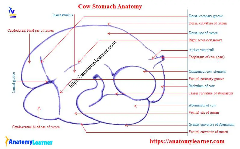
Cow anatomy stomach
It is essential to know the cow stomach anatomy as a veterinary student. The cow stomach occupies nearly three-fourths of the abdominal cavity. It fills the left half of the cavity except a small space occupying by the spleen, parts of the small intestine.
The stomach of a cow comprises four compartments – rumen, reticulum, omasum, and abomasum. I hope you know the first three parts are non-glandular, whereas the fourth part is a glandular or true stomach of a cow. The esophagus opens into a shallow depression (atrium ventriculi) between rumen and reticulum.
A cow’s rumen is in contact with the left abdominal wall from the eight intercostal spaces to the transverse plane of tuber coxae. Again, the cow’s reticulum is in contact with the left abdominal wall at the ventral end of the sixth and seventh intercostal spaces. It also contacts the ventral wall of the corresponding sternal region.
In a normal cow, the omasum is in contact with the right abdominal wall in the ventral part of the seventh to ninth intercostal spaces. In addition, it is in contact with the abdominal floor in a small area between the xiphoid cartilage and right costal cartilage.
The fundus part of the cow’s abomasum lies on the abdominal floor in the xiphoid region. Again, you will find the pyloric part of the abomasum near the ventral end of the right ninth or tenth intercostal spaces.
Compartments of cow stomach
The rumen of cow occupies most of the left half of the abdominal cavity and extends considerably to the right of the median plane. It is compressed from side to side and having two surfaces, two curvatures, and two extremities.
The parietal surface of the rumen is convex and related to the diaphragm of the cow. Again, the viscera; the surface is irregular and chiefly related to the omasum, abomasum, and intestine.
You will find right and left longitudinal grooves at both surfaces of a cow rumen. The cranial extremity divides into two sacs by the transverse groove. There are caudodorsal and caudoventral blind sacs present at the caudal extremity of the rumen.
The reticulum is the smallest, most cranial of the four compartments of cow stomach anatomy. It is somewhat piriform but compressed craniocaudally in a cow.
There is a convex diaphragmatic surface that lies against the diaphragm and liver. Again the visceral surface is flattened more or less by the pressure of atrium ruminis.
The fundus of the reticulum forms a rounded cul-de-sac that contacts the sternal part of the diaphragm, liver, omasum, and abomasum of a cow.
There is an ellipsoidal omasum that is marked off from the other compartment of the stomach. In addition, the abomasum is an elongated sac that chiefly lies on the abdominal floor. You will find three distinct portions in the abomasum of a cow.
Liver and pancreas of cow anatomy
In cow, the liver lies entirely to the right of the median plane. The long axis is directed cranioventrally from the right kidney at the last rib to the plane of the ventral third of the sixth intercostal space.
In the liver anatomy of a cow, the visceral surface is concave and possesses the essential features – porta hepatis. The right border faces caudally and is short and thick. It presents a deep impression form in the right lobe and the caudate process. There is a fossa for the gallbladder at the ventral border of the liver.
The lobulation is distinct in cow’s liver. You will find two processes in the caudate lobe of the liver.
The pancreas of the cow locates almost entirely to the right of the median plane. It consists of a large right lobe, and a small left lobe joins on the right side of the portal vein.
The left lobe encloses the dorsal attachment of the greater omentum at the root of the mesentery. In addition, the right lobe extends caudally along with the descending duodenum, enclosed between layers of the mesoduodenum.
Organs of the respiratory system from a cow
You will find a relatively short and wide larynx in the cow’s respiratory system. There is cricoid, thyroid, arytenoid, and corniculate cartilage in the anatomy of the cow larynx.
The thyroid cartilage of a cow resembles a thin, broad, cartilaginous plate that bends to form a wide U. This cartilage is relatively shorter, and its lamina is higher in goat than in the cow.
The trachea of a cow is a flexible tube that extends the caudad from the larynx at the point of the second cervical vertebrae to the level of the fifth thoracic vertebrae. You will find cervical and thoracic parts in the trachea of a cow.
The cervical part of the cow’s trachea relates with longus colli muscle dorsally. Again, the thoracic part of the trachea is in the cranial and middle part of the mediastinum.
At the left, the cow’s trachea relates dorsolaterally to the esophagus, except in the region of the tracheal bifurcation. You will find several incomplete U-shaped tracheal rings on the trachea of a cow.
Lung anatomy of a cow
Both the right and left lungs of cow anatomy occupy much of the space of the thoracic cavity. It covers by the pulmonary pleura and is free to move in the pleural sac. The right lung of a cow is almost twice as large as the left lung. This is due to the presence of an extra accessory lobe in the right lung.
You will also find a larger apical lobe in the right lung of a cow than that of the left lung. Each of the lungs contains an apex, base, two surfaces, and two borders.
The base or the diaphragmatic surface of the cow lung relates to the convex thoracic surface of the diaphragm. Again the apex of the lung occupies the space within the cupula pleurae. The apex of a cow’s right lung is much larger than that of the left lung.
There is a smooth, convex costal surface of the lung that contacts the ribs’ inner surface. The medial surface of the lung is less extensive than that of the costal surface. You will find the cardiac impression on the medial surface of the cow’s lung. The cardiac impression is much deeper in the right lung of a cow than that of the left lung.
A sharp and irregular ventral border in the lung separates the costal surface from the medial surface. The dorsal border is thick, and the basal border separates the diaphragmatic surface from the medial and costal surfaces.
The apical lobe is large and extends to the left of the median plane, ventral to the trachea. There is an elongated three-sided middle lobe present in the lung. In addition, the diaphragmatic lobe is the most prominent lobe in cow lung and caudal in position.
Heart of a cow
This is also the basic need to know the heart anatomy of a cow as a veterinarian. The fifth seventh of the heart of a cow is on the left side of the median plane. The length of cow’s heart from the base to the apex is relatively longer than that of a horse.
The base is opposite to the thoracic wall from the second intercostal space or third rib to the fifth intercostal space or sixth rib. In addition, the apex of cow’s heart is opposite the sixth chondrosternal joint. The long axis is less oblique than in the horse.
There is a more regular, conical, and more pointed ventricular part present in cow’s heart. The left ventricular border is opposite to the fifth intercostal space. A caudal groove extends from the coronary groove ventrally to the left side of the left ventricular border.
You will find large blood vessels that lodges in the coronary and interventricular groove of the cow’s heart. The left auricle of the heart is larger than the right one. A larger cardiac notch on the left side extends between the caudal border of the third rib and fifth intercostal space.
The left vena azygos opens into the right atrium ventral to the caudal vena cava. Two bones, the oss cordis, develop in the aortic fibrous ring. The right one is in opposition with the atrioventricular rings. It is irregularly triangular.
The pulmonary trunk is relatively large, leaving the right ventricle at the conus arteriosus of the cow’s heart. In addition, the right pulmonary artery lies on the right side of the median plane. This right pulmonary artery of cow’ heart is usually longer than the corresponding left artery.
Cow hoof anatomy
There are four divided hoofs on the limbs of a cow that covers the end of digits. So, the hoof anatomy of a cow is different than that of a horse. I have a detailed guide on the hoof anatomy of the horse here on anatomy learner.
You will find three surfaces on cow’s hoof – the abaxial, interdigital, and basal surfaces. The abaxial surface is convex from side to side and marks by a ridge parallel to the coronary border.
Again, the interdigital surface is concave and grooved that touches the opposite claw only at the end. In addition, the basal or the ground surface of cow’s hoof consists of two parts – the sole and bulb.
The hoof of a cow consists of three parts – peripole, wall, and sole. The peripole surrounds the coronary border, and the wall forms most of the abaxial surface of a hoof.
Cow anatomy labeled diagram
Here I would like to summarize the whole anatomical features of a cow (both internal and external) with the labeled diagram. I hope you will enjoy it and learn the anatomical features of the different organs of a cow.
If you need more cow-labeled diagrams, you may join with anatomy learners on social media.
Frequently asked questions on cow
What do you call a cow face?
Is a cow an ox?
Why choose a cow eye to study human anatomy?
Why are the sheep brain and cow eye used to study anatomy?
Why are cow hearts used to study human anatomy?
Conclusion
This is the basic information on cow anatomy for any veterinary student or farm owner. But, you might learn more about different systems of cow anatomy with a labeled diagram. , you may visit the general anatomy section of anatomy learner (guide page) and learn all the organs from different cow systems.
I always request you to practice your learning with the actual anatomical samples. You may use the materials (labeled diagrams, videos) that I provided here in this article.