The dog mandible anatomy consists of two halves of the mandibula. Here, the right and left half of the mandibles firmly unit in life at the intermandibular suture.
In this guide, I will show you the anatomical features of the dog mandible bone with a diagram. You will find the special features of the canine mandible compared to other animals, like cows, horses, and pigs.
Quick overview: the right and left halves of the mandibula form the dog mandible. It is considered the inferior jaw, and each mandible consists of a body and a ramus. The masseteric fossa and angular process are the unique osteological features of the dog mandible.
I will help you to identify all other osteological features from the canine mandible. Thus, after completing this article, you will easily identify it and differentiate it from other’s animal mandibles.
Okay, let’s start to learn the osteological features of the canine mandible with the labeled diagram.
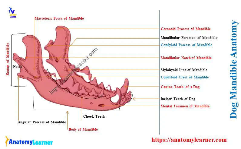
Dog mandible anatomy
First, I will show you the various osteological features of the dog mandible with the diagram. Let’s see and identify the below-mentioned features from the ramus and body of the canine mandible –
- Ramus and body of the canine mandible,
- Coronoid process (upper larger body enlargement),
- Condyloid process (lower wider bony enlargement),
- Coronoid and condyloid crests of the ramus of the canine mandible,
- The mandibular notch between coronoid and condyloid processes,
- Deep masseteric fossa on the lateral aspect of the ramus of the dog lower jaw,
- A masseteric line on the lateral aspect of the ramus of the mandible,
- Angular process of the ramus of canine mandible (caudal aspect, below condyloid process),
- Mandibular foramen (medial aspect of ramus) and mandibular canal,
- A mylohyoid line on the medial aspect of the body of the canine mandible,
- Mental foramina at the lateral cranial aspect of the body of the dog mandible,
- Alveolar sockets on the body of the mandible, and
- Different borders, surfaces, and angles of the mandible,
All these above-mentioned osteological features are identified in the canine mandible labeled diagram. I hope you will identify all these features from the real sample of dogs and differentiate them from other animal mandibles.
Unique features of the dog mandible bone
You will find the below-mentioned unique osteological features in the dog mandible bone –
- The shape of the canine mandible is somewhat less triangle in most of the breeds (compared to cows),
- You will find the incomplete fusion between two halves of canine mandibles,
- Each of the mandibles shows a larger horizontal body and a verticle ramus,
- Between the horizontal body and verticle ramus of the mandible, the mandibular angle is formed,
- Each of the body of dog mandibular bones possesses three alveoli for incisors, one alveolus for canine, and seven alveoli for molar and premolar teeth,
- You will find a notch and process (angular process) at the caudal aspect of the canine mandibular angle,
- The lateral surface of the ramus possesses a deep depression (known as the masseteric fossa),
- There are 2 larger bony projections on the upper part of the ramus–condylar and coronoid process,
- You will find the mandibular notch in between the coronoid and condylar process of the canine mandible,
- The medial aspect of each mandible bears a larger mandibular foramen that exists as the mental foramina laterally,
You should know this basic information about the canine mandible structure. But you may also learn the details of these structures of the mandible from the next section of this article.
Before that, let’s see the summary of the canine mandible’s features from Table 1 –
| Canine mandible anatomy | Unique osteological features |
| Lateral surface of ramus | Have deep masseteric fossa |
| Caudal angle of mandible | Possess angular process |
| Mental foramina | More than 2 (two) |
| Alveolar socket for canine teeth | One in each mandible |
Canine mandible anatomy description
For description purposes, you may divide the canine mandible into 2 parts –
- Horizontal body of the mandible and
- Vertical ramus of the mandible,
The body of the dog’s mandible is longer than the verticle ramus. Both the body and ramus of the canine mandible possess salient osteological features.
First, let’s discuss on the unique features of the body of the canine mandible –
Body of the dog mandible
This is the longer part of the dog’s mandible. First, let’s see the summary of the body of a dog’s mandible structure from Table 2 –
| Body of the dog mandible | Unique features |
| Parts | Two (2) – incisival and molar parts |
| Incisival part | Possess – two surfaces Concave lingual, and Convex labial surface |
| Molar part of the body | Possess – Two borders, Two surfaces, and Two extremities, |
| Dorsal / alveolar border | Thick Possess – alveolar sockets |
| Ventral border | Thick and convex |
| Medial surface of the body | Possess mylohyoid lines |
| Lateral surface | Smooth and have mental foramina |
The anterior part of the body is known as the pars incisive. In contrast, the posterior part of the mandibular body is the pars molaris.
On the anterior part, you will find the alveolar sockets (conical cavities for the roots of the teeth) for the incisor. Again, the caudal part of the body has alveolar sockets for canine, molar, and premolar teeth.
Surfaces of dog mandibular body
The anterior part also presents two surfaces –
- Convex labial and concave lingual surfaces,
Here, the medial surface of the anterior part of the body faces the tongue. Thus, it is the lingual surface,
The outer surface of the anterior or incisive part faces the lips. Thus, this is the labial part of the anterior incisive portion of the dog mandible.
The caudal part of the body has 2 surfaces, 2 borders, and 2 extremities. Here, the surfaces are lateral and medial.
The lateral surface of the posterior body faces the cheek and is known as the buccal surface. Again, the medial surface faces the lateral border of the tongue. Thus, this medial surface is also known as the lingual surface.
You may get the basic idea of the structure of the dog tongue and mouth cavity from the below-mentioned articles –
- Dog tongue anatomy with labeled diagram – muscle, papillae, glands, veins, and nerves, and
- Dog mouth anatomy – lip, cheek, oral cavity, and salivary glands with diagram,
Unique features of the dog’s mandibular body
You will find 7 alveolar sockets on the posterior body of the mandible for cheek teeth (molar and premolar). Again, there is only one alveolar socket on the molar part of the body for the canine teeth.
Thus, the total alveolar sockets on the posterior body of the mandible are – 8 (eight),
But, the total body (anterior and posterior parts) possess 3 sockets for the incisor, 1 socket for the canine, and 7 sockets for the cheek teeth.
You will find the single alveoli for the root of 3 incisors, 1 canine, and first and last cheek teeth. And the body shows 2 alveoli for the middle five cheek teeth.
The fifth cheek tooth (first molar) is the largest tooth in the canine. Here, you will find 2 important terms –
- Interalveolar margin and interalveolar septa,
The dorsal border of the mandible between the first canine and first cheek tooth is larger. This border is the interalveolar margin of the body of the dog mandible.
Again, you will see the space between the adjacent premolar teeth. This is the interalveolar septa and is very narrow in appearance.
You may know the details of the canine teeth from the below-mentioned article –
The ventral border of the body of a dog mandible is convex and thick.
Now, the medial or lingual surface of the posterior body presents a wide, smooth, longitudinal ridge. This longitudinal ridge is the mylohyoid line where the mylohyoideus muscle attaches.
Where are the mental foramina in the dog’s mandible?
The lateral surface of the body is long, smooth, and possesses a uniform width. Rostral to the lateral surface, you will find more than 2 mental foramina near the intermandibular suture.
The middle mental foramen is the largest, which locates ventral to the septum (s) between the first two cheek teeth. You will also find several smaller mental foramina on the lateral surface of the body of a canine mandible.
How is intermandibular symphysis formed in dogs?
The anterior part of the right and left mandibular body articulate firmly at the midline. Thus it forms the intermandibular symphysis in dogs between two halves of mandibles.
This intermandibular symphysis is a strong, rough-surfaced, and fibrous joint. You may know the features of the symphysis joint from the below-mentioned article of anatomy learners –
You will also find a similar type of joint (symphysis) between two halves of the hip or pelvic bones.
Ramus of dog mandible anatomy
The ramus of the mandible is the non-tooth-bearing verticle part of the bone. You will find three salient processes in the structure of the dog’s mandibular ramus –
- Dorsally – coronoid process,
- Ventral to coronoid process – condyloid process, and
- Caudal to the angle–angular process,
The caudal part of the body continues with the ventral part of the ramus of the dog mandible. Here, the caudal part of the body forms the mandibular angle with the verticle ramus.
Anatomically, each ramus of the dog’s mandible possesses –
- Two surfaces – lateral (masseteric) and medial (mandibular),
- Two borders – cranial and caudal borders, and
- 2 extremities or ends – dorsal and ventral,
The dog mandibular ramus labelled diagram identifies all these surfaces, borders, and extremities.
Coronoid process of dog mandible
The most dorsal part of the verticle ramus shows an enlarged bony projection. This is the coronoid process of the dog’s mandible, which extends dorsally and laterally.
It is a large thin plate of bone with a thickened rostral border. You will find a little difference in the appearance of the coronoid process between the dogs and cows.
Condylar process of the dog mandible
Below the coronoid process, you will find another bony process. This is the condylar process of the dog’s mandible anatomy, which faces the caudally of the ramus.
The anatomical name of the condylar process is processus condylar. This is the transversely elongated and sagitally convex articular process.
This condylar process of the dog mandible forms the temporomandibular articulation with the mandibular fossa of the temporal bone. In between the coronoid and condylar processes, you will find the notch. This is the mandibular notch of the canine mandible.
The caudoventral mandibular angle possesses a hook-like process, a more salient feature in a dog’s mandible. This is the angular process of the dog mandible structure.
You will find the attachment of the pterygoid muscle medially in this process. Again, the masseter muscle attaches laterally to the angular process of the canine mandible.
You will also find a notch between the angular and condyloid processes of the canine mandible.
Where is the mandibular fossa located in a dog?
The mandibular fossa is located on the lateral surface of the ramus of a dog’s mandible. This is a major and three-sided depression on the lateral aspect of the ramus of a canine mandible.
This well-developed and deep mandibular fossa will only be found in the canine mandible. But, the horse and cow’s mandible don’t possess well-developed and deep mandibular fossa.
Within this mandibular fossa of a dog, the masseter muscle inserts. Again, this masseter muscle also has an attachment with the rostral border of the ramus. You will also find the attachment of the masseteric muscle caudoventrally by the neck of the condylar process.
Mandibular foramen of the dog
The medial surface of the ramus of a dog mandibular bone shows less concavity. You will find the dog’s temporal muscles insertion on the medial surface of the mandible.
Just ventral to the insertion of the temporal muscle, you will find the larger mandibular foramen on the medial aspect of the dog mandible. The mandibular foramen is the caudal opening of the longer mandibular canal.
And within the mandibular canal, the mandibular nerve passes. Finally, the dog’s mandibular canal cranially opens as the mental foramina. Now, the dog’s mandibular nerve also becomes the mental nerve.
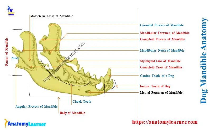
Dog mandible labeled diagram
Now, let’s see all the osteological features from the ramus and body of the dog’s mandible. Here, I tried to show all these features both from the ramus and body of the canine mandible.
The various alveolar sockets for teeth and mental foramina from the body of the dog mandible are identified in the labeled diagram. Three important processes from the ramus of the dog mandible are also identified in the labeled diagram.
The dog mandible labeled diagram also identifies the unique mandibular fossa. You may find more labeled diagrams on the canine mandible here. Or find all the canine mandible features in a video I shared here.
How to differentiate dog mandible from cows, horses, and pigs?
While comparing the dog mandible to cows, horses, and pigs, you might consider the following features –
- The appearance of the masseteric fossa on the lateral surface of the ramus,
- Presence of angular process,
- Number of alveolar sockets for teeth on the body of the mandible, and
- Number of mental foramina on the cranial aspect of the body of the mandible,
On cows, you will find the typical features of the mandible. But there are no angular processes and well-developed masseteric fossa on the ramus of a cow mandible.
You may learn more about the cow mandible from the below-mentioned article of anatomy learners –
Again, you will not find any alveolar socket for the canine teeth on the body of the cow mandible.
But, the horse mandible exceptionally shows the complete fusion between the right and left halves of the mandible. The alveolar border of the body of a horse mandible shows 6 sockets for incisor teeth.
You will also find two alveoli in the male horse mandible for canine teeth. Again, the horse mandible doesn’t show any angular or well-developed masseteric fossa.
Again, the pig mandible also shows some unique osteological features. You will see the short and pointed coronoid process in the structure of the pig mandible bone.
The angle between the two rami of the pig mandible is comparatively wider. Again, you will not find the angular process in the caudal angle of the pig mandible.
Frequently asked questions on dog mandible
Here, I will enlist the frequently asked questions on the dog mandible with concise answers. But, it is suggested to know all the information about the canine mandible I provided here.
Okay, let’s see the most commonly asked questions on canine mandible –
What is the mandibular on a dog?
The mandibular is the lower jaw bone of a dog. There are two halves of mandibles (right and left), and they fuse to form a single bone.
The body of the mandible possesses essential alveolar sockets for teeth. Again, various muscles are inserted on the lateral and medial surfaces of the ramus of the dog’s mandible.
What is the function of the mandible in dogs?
The mandible is considered the harder framework of the dog’s lower jaw. Two halves of the mandible fuse to form the intermandibular symphysis, making the lower jaw stronger.
The bodies of the dog mandible form the base of the floor of the mouth cavity where the tongue is hosted. The condyloid process of the dog mandible forms the temporomandibular articulation.
Different processes and surfaces of the dog mandible act as the insertion site for various muscles.
What muscles attach to the dog’s mandible?
Various muscles attach to the dog’s mandible. The most important muscles are the masseteric, pterygoid, temporal, digastricus, mylohyoideus, and genioglossus muscles.
You will find masseteric muscle on the lateral masseteric fossa of the dog’s mandible. Again, the pterygoid and temporal muscles are found on the medial aspect of the ramus.
The mylohyoideus muscle remains ventrally to the intermandibular articulation. The longer digastricus muscle attaches ventral to the body of the dog’s mandible.
What are the processes of the dog’s mandible?
You will find three well-developed processes in the structure of the dog mandible. All these processes are the bony extension from the ramus section of the canine mandible.
You will find the coronoid and condyloid processes at the upper aspect of the ramus. Finally, the unique angular process at the ventral mandibular angle will be visible.
Conclusion
So, the dog mandible anatomy comprises the right and left halves of mandibles. They form a strong and fibrous intermandibular articulation ventrally.
The most characteristic features of the dog mandible are the presence of deep masseteric fossa, angular process, and more than two mental foramina. You may compare the dog mandible with other animals like cows, horses, and pigs based on these unique osteological features.