The dog tarsal bones form the skeleton of the hind paw with the metatarsals and phalanges. These bones from the dog’s hind paw arrange into three transverse rows.
I will show you the number of canine tarsal bones and their detailed anatomical description.
Quick overview: 7 tarsal bones in the dog tarsus anatomy are arranged into three transverse rows. The canine tarsus is more than three times as long as the carpus. The talus and calcaneus are two well-developed and clinically important dog tarsal bones.
You will get a basic idea of the structure of the canine tarsus from this article. Again, I will show you the differences among the tarsal bones from various species like horses, dogs, cows, and pigs.
If you want to learn the anatomical features of the canine tarsal bones with a diagram, let’s continue the article until the end.
Dog tarsal bones
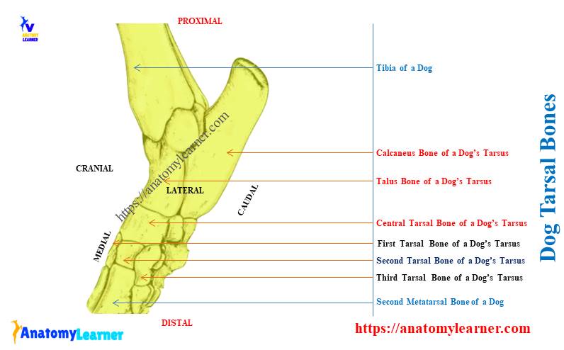
First, let’s identify the dog tarsus area from the below-mentioned labeled diagram. The canine tarsus is located between the tibia-fibula and metatarsal bones.
Here, you will find the following bones in the dog tarsus –
- Largest and longest calcaneus bone,
- Second largest talus bone,
- Central tarsal bone below the talus, and
- Tarsal I, II, III, and IV in the distal row,
Again, the talus and calcaneus bones of a dog show different osteological features. You will find the details of these osteological features of canine talus and calcaneus in the article’s next section.
Now, let’s see the summary of the dog tarsals from Table 1 –
| Canine Tarsal Bones | Anatomical Features |
| Number of the dog tarsals | 7 (seven) |
| Arrangement | Arrange in three rows Proximal row – contain 2 tarsal Middle row – contain only 1 tarsal, and Distal row – contains 4 tarsal bones |
| Name of canine tarsals | Proximal row Talus (medial), and calcaneus (lateral) Middle row Central tarsal bone Distal row Tarsal I, II, III, and IV (medial to lateral) |
| Formation of joint | They form tarsal or hock joint Tibio tarsal articulation Intertarsal articulation Tarso-metatarsal articualtion |
What are the tarsal bones in a dog?
The tarsals are the short bones in between the tibia-fibula and metatarsal of the dog’s hindlimb. These tarsal bones arrange so that the tibia and fibula articulate with the talus proximally.
Again, the distal row tarsals of a dog tarsus articulate with the proximal end of the metatarsal bones.
The canine tarsal bones are considered the short type of bone. Typically, you will find different surfaces (normally 6 surfaces) in these tarsal bones.
Again, the surfaces of the canine tarsal are irregular. Most of the short bone of a canine skeleton shows similar length, breadth, and height.
The below-mentioned article from anatomy learner might help you to get a basic idea of other different types of bone from the canine skeleton –
You may get the basic idea of the anatomy of a dog tibia from the below-mentioned article of anatomy learners –
You know, the tarsals, metatarsals, and phalanges together form the hind paw or pes in dogs. If you want to know about other different segments from the canine hindlimb and forelimb, you may go through the below-mentioned article –
Number of tarsal bones in a dog
There are seven tarsal bones in the dog’s tarsus. The length of the dog’s tarsus is more than the carpus.
Typically, the dog tarsals arrange in three rows as follows –
- Proximal row,
- Middle row, and
- Distal row,
In the proximal row, you will find the largest and longest calcaneus bone on the lateral aspect. Again, you will find the talus bone in the proximal row of the tarsal bones, which locates medially.
The middle row of the canine tarsus only possesses the central tarsal bone. Again, the distal row of the canine tarsus includes four tarsal bones.
There are three small bones – the first, second, and third tarsal bones are located side by side. They are separated from the proximal row by the central tarsal bone.
The fourth tarsal bone in the dog tarsus is the largest, which completes the distal row laterally.
Now, let’s see the arrangement of the canine tarsal bones from Table 2 –
| Canine tarsal Bones | |
| Proximal Row | Calcaneus (lateral) + Talus (medial) |
| Middle Row | Central Tarsal |
| Distal Row | First + Second + Third + Fourth Tarsals |
Hope you will identify all these tarsal bones from the real canine hind leg structure.
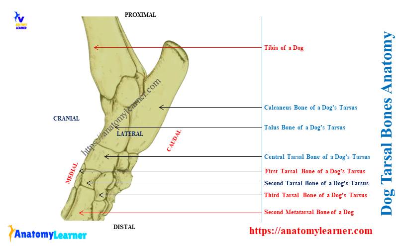
Dog tarsal bones anatomy
Now, you will learn dog tarsal bones anatomy with the labeled diagram. You might know the structure of talus and calcaneus tarsal bones as they are larger and clinically important.
Here, I will discuss the following bones from the canine tarsus (anatomical names of the dog tarsals are shown in Table – 3) –
| Canine Tarsal Bones | Anatomical Name |
| Talus (Tibial Tarsal) | Corpus tali |
| Calcaneus (Fibular Tarsal) | Os calcis |
| Central tarsal bone | Os tarsi central |
| First tarsal bone | Os tarsale I |
| Second tarsal bone | Os tarsale II |
| Third tarsal bone | Os tarsale III |
| Fourth tarsal bone | Os tarsale IV |
Okay, let’s start with the anatomy of the dog calcaneus bone.
Dog calcaneus tarsal bone
Dog calcaneus is the fibular tarsal bone in the structure of the tarsus. It is the largest and longest tarsal bone in the canine tarsus anatomy.
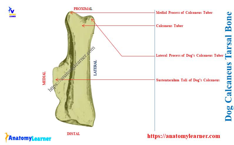
First, let’s try to identify all the below-mentioned osteological features from the canine calcaneus bone –
- Calcaneus tuber (proximally),
- Sustentaculum tali (just above the articular surface for talus),
- Articular surfaces for the talus bone, and
- Body of the calcaneus bone,
The labeled diagram identifies all these features from the dog calcaneus tarsal bone.
The distal half of the dog calcaneus bone is wide transversely. You will find three articular surfaces for the talus bone on the distal half of the calcaneus bone.
There are also two processes (bony elevation) in the structure of the canine calcaneus bone. Proximally, you will see the larger bony process, which is known as the calcaneus tuber.
This calcaneus tuber serves for the insertion of the common calcaneus tendon. This (common calcaneus tendon) is the insertion part of the gastrocnemius, gluetiobiceps, and semitendinosus muscles that insert on the calcaneus tuber.
In the structure of the calcaneus tuber of a dog calcaneus bone, you will find two processes – lateral and medial processes. A wide groove separates these lateral and medial processes on the tuber calcaneus.
On the plantar surface of the calcaneus tuber, you will see a wide but shallow groove. Within this groove, the tendon of the flexor digitorum lateralis muscles runs.
Articular surfaces on dog calcaneus bone
There are three articular surfaces for articulation with the tibial tarsal or talus bone. The dorsomedial articular surfaces are concave oval, and the lateral articular surface is convex.
You will also find a very small articular surface for the articulation with the central tarsal bone. The calcaneus sulcus locates between the middle and distal articular surfaces.
Again, the distal surface of the calcaneus bone shows a larger flat articular surface. This large articular surface is for articulation with the fourth tarsal bone. A small articular surface of the talus bone also designs for articulation with the fourth tarsal bone.
The articular surfaces from the canine calcaneus bone are also identified in the labeled diagram.
Dog talus bone – fibular tarsal
The talus of a dog is the second largest tarsal bone. This bone articulates proximally with the tibia and fibula, distally with the central tarsal. You will also find the articulation of the distal part of the dog talus with the plantar aspect of the calcaneus bone.
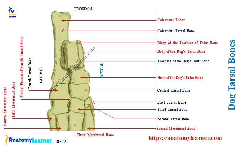
First, let’s identify the below-mentioned osteological features from the canine talus bone –
- Body of the talus bone
- Neck and head of the dog’s talus bone,
- Lateral process of the canine talus bone, and
- Trochlea of the dog talus bone,
Thus, for the description purpose, the dog talus bone may divide into three major parts – head, neck, and body. Here, the body of the talus forms the proximal half of the bone.
The trochlea is the most important osteological feature of the body of the canine talus. This trochlea possesses a middle groove with two lateral semicircular ridges.
This structure attaches to the sagittal groove of the intermediate ridge of the distal tibia bone. Again, you will also find the articulation of the two lateral aspects of the trochlea with the lateral and medial malleoli bones.
On the plantar surface of the talus bone, you will also find the articular surfaces for the calcaneus. There are three distinct articular surfaces for articulation with the calcaneus bone.
You will find the large and concave articular surface on the plantolateral surface of the dog talus bone. Again, the larger process on the lateral aspect of the talus bone is known as the lateral process of the talus bone.
An oval middle articular surface is between the lateral and deep parts of the talus bone. You will find the smallest articular surface for the calcaneus bone at the distolateral part of the dog talus bone.
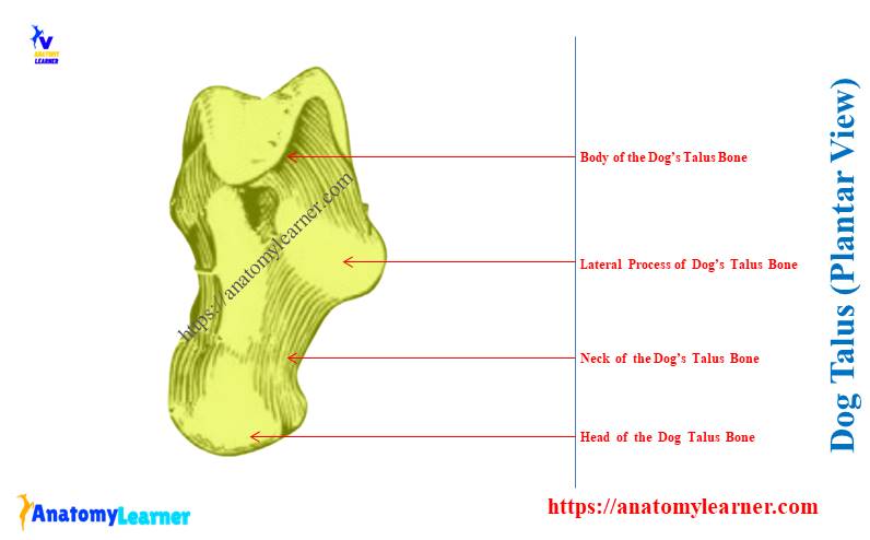
Head and neck of the dog’s talus bone
The head of the dog’s talus locates on the distal part of this tarsal bone. This is the transversely elongated structure in the dog tarsus anatomy.
The anatomical name of the head is caput tali, and it possesses a distal surface. This distal articular surface on the head is a rounded and irregularly oval structure. You may call it the articular surface for the central tarsal.
No other articular surfaces are present in the distal extremity of the head of the dog talus bone.
The neck is large and located between the head and body of the canine talus bone. It is smooth and convex medially (m) and lies directly adjacent to the skin of the dog’s leg.
Canine central tarsal bone
The central tarsal is the only bone in the middle row of the canine tarsus. It lies medially and locates between the proximal and distal rows of the canine tarsal bones.
You will find the articulation of the canine central tarsal bone with other tarsals. Proximally, you will find the large, concave, roughly oval area on the central tarsal. This articular surface of the canine central tarsal articulates with the talus.
Let’s see the articulation of the central canine bone with other tarsals –
- Proximally – with talus tarsal bone,
- Proximal plantar surface – articulates with calcaneus bone,
- Distally (medially) – articulate with the first, second, and third tarsal bones, and
- Distally (laterally) – articulates with the fourth tarsal bone,
On the proximal extremity of the dog’s central tarsal bone, you will find the tuber planteris process on its plantar aspect. In this process, you will find a small articular surface for articulation with the calcaneus tarsal bone.
Again, the central tarsal bone articulates with the first, second, and third tarsal bones distally. Finally, the lateral aspect of the central tarsal (c) articulates with the proximal half of the fourth tarsal bone.
Dog first, second, third, and fourth tarsal bones
The distal row of the dog tarsus shows 4 tarsal bones – first, second, third, and fourth. You will find great variation in the development of the first tarsal bone in a dog.
Sometimes in dog legs, you find this first tarsal as the fused bone with the first metatarsal. So when fuses with the metatarsal, you will find this first tarsal as the rough and bent plate-like bone.
But when it exists as the separated bone in the dog tarsus, you will see the transverse compression. Let’s see the typical articulation of the first tarsal bone with other bones –
- With central tarsal bone,
- Second tarsal and first metatarsal bones,
Sometimes, you may find the articulation of the first tarsal bone with the second metatarsal bone.
The dog’s second tarsal is the smallest bone in the tarsus anatomy. It is a wedge-shaped bone that extends toward the plantar aspect of the hind paw.
Let’s see the articulation of the dog’s second tarsal bone with others –
- Proximally – articulates with the central tarsal bone,
- Laterally – articulates with the third tarsal bone,
- Medially – articulates with the first tarsal bone, and
- Distally – articulates with the second metatarsal bone,
Canine third and fourth tarsal bones
The canine third tarsal is three times larger and two times longer than the second tarsal bone. You will find the various articulations of the dog’s third tarsal bone with others as follow –
- It proximally articulates with the central tarsal bone,
- Distally with the third metatarsal bone,
- Laterally with the fourth tarsal bone,
- Medially with the second tarsal and metatarsal II bones,
You will find a rounded plantar tuberosity on the planter aspect of the third tarsal bone.
The fourth tarsal is the longest and largest bone in the distal row of a dog’s tarsal. Centrally, you will find a medial process in the structure of the canine fourth tarsal bone.
You will find the following articulations of the dog tarsal bone with others –
- The canine fourth tarsal articulates with the central and third tarsal bones medially,
- Proximally articulates with the calcaneus and also with the talus bone dorsomedially,
- Distally articulates with the metatarsal IV and V bones,
Here, the articulation between the fourth and central tarsal slopes proximally and laterally. Again, the between the fourth and third tarsal slopes distally and medially.
You will see a wide groove on the distal half of the lateral surface of the canine fourth tarsal bone. This wide groove is designed to pass the tendon of the canine fibularis longus muscle.
Proximal to this groove, you will also find the salient tuberosity of the canine fourth tarsal bone. Again, two indistinct rectangular areas are on the distal extremity of the dog’s fourth tarsal bone.
Dog tarsus anatomy
The dog tarsus anatomy consists of those mentioned above 7 tarsal bones. The term “dog tarsus” also means the collection of several joints among the tarsal, metatarsal, and tibia-fibula bones.
Here, the tarsal bones of the canine tarsus anatomy form the tibiotarsal joints with the tibia bone. In between the tarsal bones of the canine tarsus, they form intertarsal articulation.
Finally, the distal end of the tarsal bones forms the tarsometatarsal articulation with the proximal ends of the metatarsal bones.
Collectively, all these joint of the dog tarsus is known as the hock joint. You may get the full idea of canine hock articulation from the below-mentioned article of anatomy learners –
Tarsal bones of horse
You will find 6 (six) tarsal bones in the horse tarsus anatomy. Again, these horse tarsal bones arrange in three rows – proximal, middle, and distal.
You will find the talus and calcaneus bone in the proximal row of the horse tarsus. Again, the middle row of the tarsal consists of only the central tarsal in horses.
Finally, you will find 3 (three) tarsal bones in the distal row of the horse tarsus. From the lateral to the medial aspect of the distal row, you will find the followings –
- Fourth tarsal bone (below the talus),
- Third tarsal bone, and
- Fused first and second tarsal bones (medially),
The tibial tarsal and fibular tarsal of the horse tarsus are short and thicker compared to the dogs and cows. You will find the oblique groove at the proximal extremity of the talus bone of a horse.
The horse’s central and third tarsal bones are flat in shape. Again, the first and second tarsal of the horse are fused and small. But the fourth tarsal of the horse is larger and irregular.
How to differentiate dog tarsals from cow tarsal bones?
You will find only 5 developed tarsal bones in the structure of a cow tarsus. The well-developed talus and calcaneus tarsal bones are in the proximal row.
Again, the middle row of the cow tarsal shows the fused central and fourth tarsal bones. Here, you will find the difference between the tarsal bones between dogs and cows.
The distal row of the cow tarsal also possesses fewer bones (only 2) than the dogs. You will only find the first and fused second and third tarsal bones in the structure of the distal row tarsal of a cow. But, in the case of a dog, you will find 4 tarsals in the distal row of the tarsus.
You will also find the variation in the structure of the tarsal bones between cows and dogs.
Frequently asked questions on the dog tarsal bone
Let’s see the commonly asked questions on the dog tarsal bone that the learners frequently ask. But, you might have the basic idea of the structure of every single bone from the canine tarsal anatomy.
Okay, let’s see the questions on the canine tarsal bone with their concise answer –
How many tarsal bones does a dog have?
Answer: there are 7 tarsal bones in a dog’s tarsus anatomy that arrange into three rows. The proximal row of the canine tarsal shows medial talus and lateral calcaneus bones.
The middle row shows only the central tarsal bone in the dog tarsus. Again, the distal row of the dog tarsal possesses 4 well-developed tarsals – first, second, third, and fourth.
What are the 7 tarsal bones?
Answer: the 7 tarsal bones are – talus, calcaneus, central, first, second, third, and fourth. Together, they form the hock joints when they articulate proximally with the tibia-fibula and distally with the metatarsal bones.
Again, these 7 tarsal bones of the canine hind leg contribute to the pes.
What do tarsals do in dogs?
Answer: the tarsals form the hock joint in a dog. Again, the tarsal is the main component of the pes structure in a dog.
Different osteological features of the tarsal bones are designed to attach various ligaments and tendons. Finally, it works as the framework of the pes and the basis of the passing of multiple muscles.
Conclusion
So, the dog tarsal bones are seven in number and arranged in three rows in the tarsus anatomy. To understand the hock joint anatomy, you might know the structure of these tarsal bones from dogs.
The dog’s tarsal bones form the hock and pes in its hind leg. Now, you might identify all these canine tarsal bones from the real sample of tarsus.