If you want to learn cartilage histology, you might know about the histology of hyaline cartilage, elastic cartilage, and fibroealstic cartilage. I have published an article related to the histological features of hyaline cartilage previously. Today, I will describe elastic cartilage histology with slide images.
Hello, welcome back again to anatomlearner, and thank you so much for getting into this short but important article. This article is the continuation of learning the histological features of cartilage.
I will show you every single histological feature of elastic cartilage from real histological slide pictures. Again, I will also provide you the identification points of elastic cartilage histology slide under the light compound microscope.
After reading this short article, you will able to identify the most important structure from the elastic cartilage histological slide.
The labeled diagram of elastic cartilage histology will help you understand the basic characteristics of elastic cartilage properly.
If you are interested in learning elastic cartilage features, then continue this article till the end.
Elastic cartilage histology
First, you should know the important structures of elastic cartilage that you might identify under the light compound microscope. Well, here I will enlist the most important features or structures from the elastic cartilage histology slide.
- #1. Perichondrium of elastic cartilage
- #2. Fibrocytes in the perichondrium of elastic cartilage
- #3. Chondrogenic layer of perichondrium of elastic cartilage
- #4. Lacunae with larger and smaller chondrocytes within the matrix of elastic cartilage
- #5. Elastic fibers (thinner and thicker fiber; most important features)
- #6. Cartilage matrix with elastic fibers
- #7. Nuclei of small and large chondrocytes with the matrix of elastic cartilage
- #8. Stratified squamous epithelium (if epiglottis)
- #9. Lamina propria (if the sample is epiglottis)
Okay, let find out these histological features from the sample elastic cartilage microscope slide.
Identification of elastic cartilage slide
If you want to identify the histology slide of elastic cartilage, the following important identifying characteristics will help you a lot. You may enlist other identifying characteristics of elastic cartilage slide if you want.
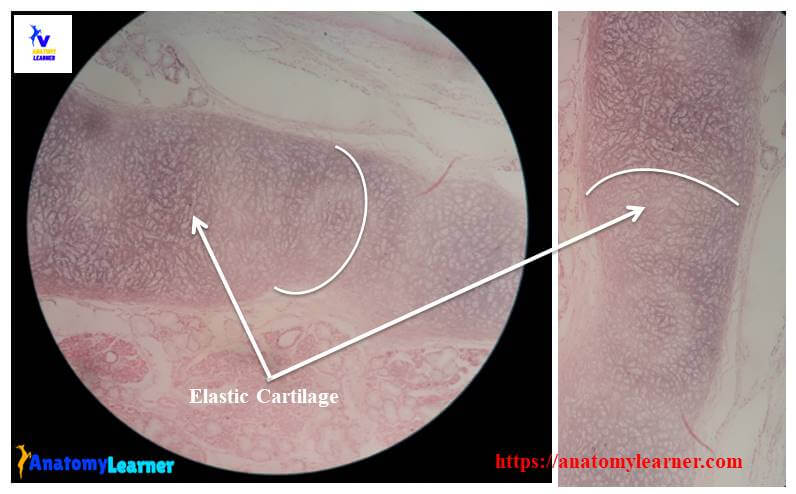
#1. The sample tissue section shows numerous thin, branching, and anastomosing elastic fibers within the matrix (matrix of elastic cartilage)
#2. Presence of closely packed chondrocytes with eccentric nuclei in the lacunae of matrix
#3. In the center of the sample tissue (within the matrix) shows larger chondrocytes in the lacunae
#4. The smaller chondrocytes located in the periphery of tissue matrix
#5. Presence of perichondrium that covers the sample tissue (matrix)
So, this is a slide of elastic tissue structure.
Elastic cartilage histology description
Well, now I will describe the elastic cartilage histology details with labeled diagram and slide images. You might find the same components in elastic cartilage that you have found in the structure of hyaline cartilage.
In elastic cartilage histology, you might describe the cells of elastic cartilage, fibers, and ground substance of elastic cartilage (matrix of cartilage).
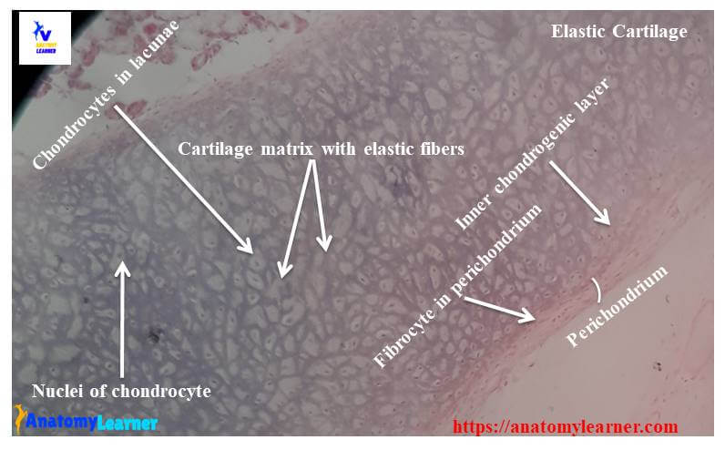
Cells of elastic cartilage structure
You will also find the same chondrocytes in the lacunae of the elastic cartilage’s matrix. In the central lacunae of matrix, you will find the larger chondrocytes and smaller, flatten chondrocytes peripheral of matrix. Chondrocytes located in the center of the elastic cartilage structure contain numerous fat vacuoles. These fat-containing chondrocytes become adipose tissue later.
Matrix of elastic cartilage histology
The matrix of elastic cartilage contains the fibers and ground substances. You will find numerous elastic fibers in the extracellular matrix of elastic cartilage. These elastic fibers enter into the elastic cartilage matrix from the surrounding connective tissue perichondrium.
Elastic fibers of elastic cartilage histology slide are thin and form a branching and anastomosing network visible with routine histology staining. In the same cartilage, the density of these elastic fibers varies among different areas. You will find a few elastic fibers near the perichondrium of elastic cartilage.
Perichondrium of elastic cartilage
The histological structure of the perichondrium is almost similar to the perichondrium of hyaline cartilage. Here, you will also find the two distinct layers – the inner chondrogenic and outer fibrous layers. You know the chondroblast develops from the inner chondrogenic layer of perichondrium and form the cartilage matrix.
You will also find the same histological features in the perichondrium’s outer fibrous layers that you found in the hyaline cartilage. There are connective tissue fibroblast and blood vessels in the outer layer of the perichondrium of elastic cartilage histology.
Location of elastic cartilage
It is also important to know where you will find the elastic cartilage in the animal’s body. I will provide some examples of organs or structures where you will find the elastic cartilage.
- #1. Structure in epiglottis (maybe the best example)
- #2. Ear pinna structure
- #3. External auditory meatus histology structure
- #4. Histology of auditory tube
- #5. In the corniculate and cuneiform cartilage of animals
Elastic cartilage histology labeled diagram and drawing
I hope these elastic cartilage slide images were helpful for you to understand the histological features of elastic cartilage. Now I will also provide the elastic cartilage histology labeled diagram for better understand.
If you need more real elastic cartilage slide images, you may follow the anatomyleaner blog on social media for fast updates.
Hyaline cartilage structure
You may read the details guide of hyaline cartilage if you want to differentiate elastic cartilage’s histological features from hyaline cartilage microscope slide. You might also interest in knowing the fibrocartilage histology from anatomy learner.
Don’t miss any single article publish here in the anatomy leaner blog. You might read other different articles related to veterinary anatomy and histology from anatomyleaner –
#1. Histological features of connective tissue cells with labeled diagram
#2. Histology of muscular tissue (smooth muscle, cardiac muscle, and skeletal muscle histology)
Function of cartilage
The functions of hyaline cartilage are –
#1. Provide support the soft tissue of the animal’s body
#2. Hyaline cartilage allows the tissue to bear mechanical stress and allow free movement of the joints
#3. This is also a shock-absorbing and sliding area for joints and facilitates the bone movements
These are the important functions of elastic cartilage –
#1. Elastic cartilage provides the strength and maintains the shape of the structure
#2. It also helps to change the shape of the structure where necessary
The most important functions of fibrocartilage are –
#1. It can resist deformation under great stress
#2. Fibrocartilage is important in attaching bone to bone and providing restricted movement
Conclusion
I think this is the best guide to know elastic cartilage histology online. This guide might help you to identify the elastic cartilage microscopic slide under the light compound microscope.
You may share this article with your friend who wants to learn elastic cartilage histology with labeled diagram and with real slide images.
Don’t forget to join social media of anatomy leaner to get fast updates on articles and different labeled images.