Veterinary students and farm owners might know the external and internal goat anatomy to maintain good health and increase the productivity of a herd. I will discuss the external and internal anatomical facts of a goat here in this article.
This brief outline of the goat’s anatomy is a starting point for those who want to keep the goat. Again, this guide is also essential for the established farm owner if they like to fine-tune their knowledge of goat anatomy.
I will try to discuss the basics of goat anatomy skeleton, internal organs, goat anatomy muscles, and more that you might know. In addition, you will find more information on anatomical features of different organs system here in this caprine anatomy learning section.
External anatomy of goat
Okay, It would be best if you had a basic idea of the external anatomy of a goat. It will help you to evaluate a goat for your herd. All goats selected for your herd need to be structurally sound.
There are many factors in evaluating a goat for replacement herd, sales, or dispersal. If your primary purpose of goat keeping is to provide meat, the amount of muscling becomes essential. Muscling should be apparent throughout the body of a goat. Again, if you want to keep a goat for wool production, you might understand the fiber types and quality of a goat.
Here, I will show you the different external anatomical features of a goat. I hope the following labeled diagram of external goat anatomy might help you get a basic idea of their body parts.
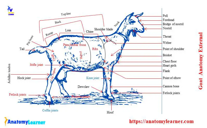
For easy understanding of the external features of a goat body, it divides into five major regions –
Head region of a goat – consists of a mouth, horns, head crest, forehead, face, poll, checks, chin, bridge of the nose, eye, and ear.
The Neck Region of a goat – consists of the neck crest, dewlap, brisket, and jugular groove.
A goat’s body or barrel consists of a hump, wither, back, loin, rump, pin bone, heart girth, ribs, flank, tail, switch, and others.
Forelimbs or forequarter of goat – consists of shoulder, arm, forearm, knee joint, cannon bone, fetlock, pastern, coffin joints, hoof, etc.
Hind limbs or hind quarter consists of the hip-joint, stifle joint, thigh bone, leg bone, hock joint, Achilles tendon, and others.
Make sure you know and identify all the external features from these five regions of the goat body.
Know more on external features of goat
You will find a head crest in between the base of the goat’s horn. There is a forehead that locates below the head crest and above the level of two eyes. The forehead of a goat is flat, bulging in shape.
The poll of the goat’s head is a bulging portion locates at the top middle center of the forehead. There is a fleshy and pink color chin below the lower lip of a goat.
The neck crest is the upper portion of the neck between the head crest and the point of wither of goat.
You may find a hanging loose, wavy fold of skin (dewlap) between chain and brisket in some goat breeds. The dewlap is more developed in cattle.
The brisket is a fleshy bulging mass between and in front of the forelimbs of the goat. You might find a jugular groove running down the neck above the trachea of a goat.
There are different essential features of the external anatomy of a goat in the body or barrel region. But I would like to talk on the back, loin, rump, and flank of a goat body.
The back of the goat’s body is a portion between the point of the wither and the last ribs. You will find a triangular area formed by joining the head of the last ribs to the two-pin bones. This triangular area is known as the loins of the goat.
Again, the rump is also a part of the back and runs from the hip-bone to the end of the goat body. The heart girth is the circular measurement around the cheast of a goat.
There is a triangular depression area (flank) of the abdominal cavity between the last rib and os-coxae bones.
Identification of a healthy goat
Sometimes you need to identify a healthy goat for your herd or other purposes. The following external appearances might help you to identify a healthy goat.
- The goat doesn’t limp or bend it back while standing or walking.
- You will find a slightly dry nostril in a healthy goat. The eye and nostril are not runny or have excessive mucous.
- The eyeball of the healthy goat will be clear and shiny.
- It doesn’t have missing hair. And the hair of the healthy goat will look smooth and shiny.
- The mucous membrane will be pink, not white, of a healthy goat.
- It breathes easily and doesn’t paint.
- The healthy goat moves with the herd or herd of the other animals.
- It eats feed in normal quantities and chews its cud.
- You will not find any bloated stomach in a healthy goat.
Thus you may identify a healthy goat with its external appearances. You may also consider the body condition, muscle attachment, and other different features to identify a healthy goat.
General goat anatomy
Here, in general, goat anatomy part, I will discuss the different organs systems of a goat. You will get a piece of basic knowledge on the goat skeleton, different types of joints, anatomical features of digestive organs, cardiovascular organs, respiratory organs, and more.
You will find many peculiar features in the different organs systems of a goat compare to other mammals. If you want to learn the detailed anatomical features of goats from different organ systems, you may read all the sheep and goat section articles.
You should not avoid learning the different bones, muscles, nerves, critical internal organs like stomach, heart, lung, intestine, and others from a goat.
Great, you will find different types of bones in the skeleton of goat-like other mammals. I have a complete guide on the skeleton of a goat and also on the muscles of a goat.
Why will you learn goat anatomy?
If you are a veterinary student, you might learn the goat anatomy for the following purposes. The farm owner might also know the minor internal anatomical features of a goat to fine-tune their knowledge.
As a veterinarian, you might practice in the field. Sometimes, you may handle the cases of a goat. First, you should perform a general examination of the goat. For that, you might have a piece of good knowledge of both the external and internal anatomical features of that goat.
Suppose you want to take the respiration rate, heart rate, impulse of the goat. Then you might know the exact location of the lung, heart, and vein of a goat. If you want to collect the rumen or abomasum fluid, you might know their particular anatomical location. Without knowing their exact anatomical location, you will not collect their fluid.
Again, if you want to perform a surgery or administration on any particular area of the goat’s body, you might know the anatomical facts of that area. Why should you know the specific anatomical facts of that area? It is necessary to avoid unnecessary injury to some essential parts, nerves, arteries, muscles, etc.
Goat Skeleton (skeletal system)
As for all domestic mammals, the skeletal system of a goat consists of the axial and the appendicular skeleton. You will find the skull, vertebral column, ribs, and sternum in the axial skeleton of a goat. At the same time, the appendicular skeleton of a goat consists of forelimbs and hindlimbs bones.
The shoulder girdle of the forelimb of a goat consists of the scapula, coracoid, and clavicle. You will find a single large bone (humerus) in the arm of a goat. In the forearm of a goat, there are two bones – radius and ulan. The maneus of a goat consists of carpus, metacarpus, and digits.

The pelvic girdle consists of os coxae, sacrum, and the first three or more coccygeal vertebrae. You will find a single bone in the thigh (femur) region of the goat that articulates the acetabulum above and tibia below. The leg bone of a goat consists of the tibia, fibula, and patella. Again, in pes of goat leg, you will find the tarsus, metatarsus, and digits.
There are a group of flat and irregular bones find in the skull of a goat. The occipital, sphenoid, and ethmoid are the single bones present in the cranium of goat’s skull. In addition, you will find a single bone (hyoid and vomer) at the face region of the goat skull.
There are seven cervical, thirteen thoracics, six or seven lumbar, four sacrum, and thirteen to nineteen coccygeal vertebrae in the vertebral column. I have described all the peculiarities of the anatomy of the goat skeleton in my previous article.
You will find thirteen pairs of elongated, curved ribs between the thoracic vertebrae and sternum of a goat. In addition, there is six unpaired segments (sternebrae) present in the sternum of a goat.
Joints of goat
The joint of a goat bone is formed by the union of two or more bones or cartilages by some binding materials and is responsible for the body’s movement. You will find three major types of joints in the skeleton of a goat – synarthrosis, diarthrosis, and amphiarthosis.
Don’t forget to learn the structure of a synovial joint if you are a student. I will enlist the different joints from the axial and appendicular skeleton of a goat.
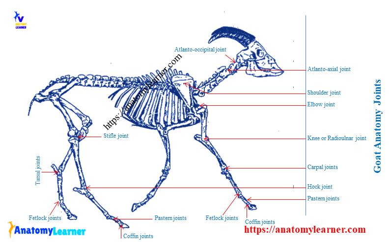
You will find the following important joint in the appendicular skeleton of a goat –
- Shoulder or scapula-humeral joint of goat
- Elbow joint of goat
- Carpal or knee joint of a goat skeleton
- A fetlock, pastern, coffin joints in the forelimb of a goat
- A sacro-iliac joint of a goat
- Symphysis joints in between two parts of oss coxarum of a goat
- Stifle joint (femoro-patellar and femorotibial joints) of goat
- Tarsus joint or hock joint of a goat
- Fetlock, pastern, and coffin joints in the hindlimb of a goat
Again, you will find the following vital joints in the axial skeleton of a goat –
- Mandibular symphysis of goat mandible
- A temporomandibular joint of goat
- Atlanto-occipital joint of goat
- An atlanto-axial joint of a goat
- Intercentral and interneural joint of the vertebrae of goat
- A costo-central joint of ribs and vertebrae of a goat
- Costo-transverse, costochondral, and costo-sternal joint in goat skeleton.
But, if you want to learn the detailed anatomical features of goat’s joint, you may read the article from the joint anatomy section.
Goat anatomy muscles
In goat muscles anatomy, I will show you some essential muscles and their structures with labeled diagrams. Ensure you know the basic anatomical terms related to muscles like– origin, insertion, tendon, and aponeurosis.
There are many muscles in the body of a goat that I tried to show you with the labeled diagrams. But here, you will know some of the essential muscles that you need in-field practices.
The goat’s masseter muscle originates from the maxilla, the fascial crest of the zygomatic bone, and the ventral surface of the zygomatic arch. This muscle inserts on the lateral surface of the ramus of the mandible of the goat.
The brachiocephalic muscles of a goat’s neck region have two parts, and superficially it relates to the skin and fascia. At the musculospiral groove of goat’s humerus, you will find the brachialis muscle. Superficially, it connects with the long and lateral head of the triceps brachii muscles of the goat.
The biceps femoris, middle glutes, semitendinosus, and semimembranosus are the essential superficial muscles in the thigh region of a goat. The semimembranosus muscle of the goat originates from the ventral surface of the ischiatic tuber and inserts at the medial condyle of the goat’s femur.
You will find two heads of gastrocnemius muscles of goat that insert a powerful, combined tendon to the calcaneal tuber. In addition, you might have a piece of good knowledge of the four abdominal muscles of a goat if you want to perform any surgeries on the abdomen.
Some other essential muscles of goat – ongissimus dorsi, longissimus costarum, intercostal extreni, and intercostal interni.
Goat anatomy internal organs
You might have good knowledge of the anatomy of the internal organs of a goat for field practices. The most important internal organs of a goat are the stomach, liver, heart, lung, intestine, etc. There are some other internal organs or parts of a goat that were described in the different articles.
You will find a compound stomach in the goat digestive system, whereas a simple stomach is present in a dog or cat. In addition, there are U-shaped incomplete cartilaginous rings in the trachea of a goat. Again, there is some variation in the larynx’s cartilage in a goat compared to the other mammals.
The structure of the goat’s heart is almost similar to that of the cattle. But, you may find some variation in the distribution of nerves and arteries of goats compared to cattle.
Now, I will discuss some essential organs or parts from the digestive, cardiovascular, respiratory, and other different goat systems.
Digestive organs of a goat
The apparatus of the goat digestive system consists of two divisions (alimentary canal and accessory organs). You will find the mouth, pharynx, esophagus, small and large intestine in the alimentary part of the goat digestive system. Again, the accessory division consists of the tongue, salivary glands, liver, and pancreas.
The mouth of goat is the first part of the goat digestive system used for holding, grinding, and mixing feed. There are two musculo-membranous folds (lips) that surround the orifice of the goat’s mouth. You will find a narrow tongue locates on the floor of the mouth cavity, between the rami of the mandible.
There is filiform, fungiform, lenticular, and vallate papilla on the surface of the goat tongue. But, you will not find the foliate papilla on goat’s tongue.
The pharynx of a goat is a musculo-membranous sac that forms the common passages for both respiratory and digestive systems. There are three parts in the pharynx of a goat. You will find seven openings on the pharynx of goat as like cattle.
The esophagus of the goat is a tube that extends from the pharynx to the stomach. You will find three parts in the esophagus of goat- cervical, thoracic, and abdominal regions.
The stomach of a goat
The stomach of a goat is a significant dilatation of the alimentary canal just behind the diaphragm. It consists of four compartments and covers almost all the left half of the abdominal cavity of the goat. The rumen, reticulum, and omasum are collectively known as the forestomach of a goat. Again, the fourth compartment that is abomasum is the actual stomach of the goat.
I have a detailed guide on ruminant stomach here on anatomy learner. If you want to know more about the ruminant stomach, you may read that article.
The rumen is the most significant part of the forestomach of a goat and consists of two surfaces, two extremities, and two curvatures. Do you know which compartment is located most cranially? Well, the reticulum is the most cranial part of the forestomach of a goat. The folds of the present reticulum honey-comb-like appearance in the interior surface.
In addition, the omasum lies to the right of the ruminoreticulum, just caudal to the liver. It is not in contact with the abdominal wall in the goat. Internal to the goat’s omasum, you will find lamina omasi.
A abomasum is the fourth compartment of the goat stomach that is the actual glandular part. You will find three different regions (cardiac, fundic, and pyloric) at the interior portion of the goat’s abomasum.
The intestine of goat anatomy
The small intestine of goat anatomy consists of three different parts – duodenum, jejunum, and ileum. In addition, you will find three other parts in the large intestine of the goat.
The duodenum is the most widespread, shortest, and fixed part of the small intestine of a goat. You will find an S-shaped curved in the duodenum of a goat.
You know a bile duct and the pancreatic duct join together and open at the same portion of the duodenum, just away from the pylorus.
In addition, the jejunum is the longest part of the small intestine of a goat. You will find a short and last part of the small intestine (ileum) joining the large intestine.
Accessory organs of goat digestive system
I hope you know the liver is the largest gland of the goat’s body, red-brown and quite brittle. It locates immediately caudal to the diaphragm and tends to be found on the right slide of the abdomen.
The liver of the goat presents two surfaces – the parietal and the visceral surfaces. You will find some ribs’ impression on the parietal surface of the goat liver.
On the visceral surface of the goat liver, you will find the impression of the following organs –
- Reticular impression
- Osmasal impression on goat liver
- Abomasal impression on goat liver and
- Renal impression on goat liver.
You will find a pear-shaped sac that lies partially in contact with the visceral surface of the goat’s liver. A soft, oyster-shaped spleen lies on the dorsal surface of the rumen just behind the diaphragm. The pancreas of the goat lies to the right of the median plane, on the visceral surface of the soft liver, and attached to the duodenum.
The respiratory system of goat anatomy
The respiratory system of goat anatomy consists of two categories of anatomical structure- the respiratory passages and the essential organs. You will find a nostril, nasal cavity proper, and conchae in the nasal cavity of a goat.
The pharynx is the second part and is very important in the goat respiratory system. You will find the paired and unpaired cartilage, muscles, and mucosal membrane in the larynx of a goat.
The trachea of a goat is a flexible, cartilaginous, and membranous tube that passages of air to and from the lung. It is kept permanently open by a series of incomplete cartilaginous rings embedded on its wall.
The diaphragm of a goat is a broad, unpaired, dome–shaped muscle that forms a partition between the thoracic and abdominal cavities. You will find three attachments on the diaphragm of a goat –
- Dorsally- the vertebral attachment,
- Laterally – the costal attachment and
- Ventrally – the sternal attachment.
The diaphragm of the goat, while separating the thoracic and abdominal cavities, gives passages to the number of structures passing both directions. You will find three passages (hiatus) on the goat diagram for the aorta, esophagus, and caudal vena cava.
Lung of goat
The lungs are the pair of respiratory organs that cover the central part of the thoracic cavity of a goat. They are covers with the pulmonary pleura. The right lung of a goat is larger than the left one.
A goat’s lung is usually pink in color and soft, spongy, and highly elastic in appearance. You will find an apex and the base in goat lung as found in cattle or other mammals.
Each part of the goat’s lung consists of two surfaces and three borders. In the right lung of a goat, you will find four different lobes – apical, cardiac, diaphragmatic, and accessory lobes. But in the left lung of a goat, there are apical and diaphragmatic lobes. The diaphragmatic lobe is the most prominent in the lung of a goat.
Frequently asked questions on a goat.
Again, in this part of the article, I will try to cover the most commonly asked questions on goat anatomical features. If you don’t find your desire question on the anatomical features, please let me know.
What is the body structure of a goat?
In a goat, you will find a skeleton that consists of both axial and appendicular parts. The axial skeleton of the goat consists of skull bones, vertebrae, ribs, and the sternum. In addition, the appendicular skeleton consists of the bones from the forelimbs and the hindlimbs.
If you want to know the details body structures of a goat, please read the full article from the sheep and goat section.
What are the internal parts of a goat?
There are lots of internal organs of a goat. Some of these organs are very important to know as they require field practices, diagnosis, and treatment. You should know the detailed anatomy of the internal organs of a goat-like stomach (four different parts), intestine, heart, lung, and other.
You will get the complete guide on the different internal organs here in this anatomy learner. Please go to the all guide section and find your desire organs systems if you wish to know.
How many stomachs do a goat have?
The goat has only one compound stomach. You will find four different compartments in the compound stomach of a goat. The four compartments of the goat stomach are the rumen, reticulum, omasum, and abomasum.
The rumen is the largest compartment of these four parts of the stomach. There is a complete guide on goat stomach here in anatomy learner.
What are the four compartments of goat?
I hope you got your answer already. Okay, the four compartments of a goat’s stomach are the rumen, reticulum, omasum, and abomasum.
You might know the topographic location of these four compartments of the goat stomach for practical purposes.
If you want to get the labeled diagrams of other different organs systems of a goat, you may join with anatomy learners on social media.
Conclusion
So, in this article, I tried to discuss all the essential information to get the basics of goat anatomy. Now, you may try to learn the details anatomical facts of a goat’s external and internal organs. You may learn both goats’ internal and external anatomy with an anatomy learner, or you may go with the books.
Please use the original resources to practice the different anatomical features of a goat. I hope the labeled diagrams of a goat’s organs systems might help you reach your destination.