Intramembranous ossification is the process of bone formation directly from mesenchyme. In this guide, I will discuss the steps or process of intramembranous ossification with a labeled diagram.
If you are interested in learning the process of intramembranous ossification in detail, you may continue this guide.
You will also know about the term – ossification center, osteoid, and mineralization here in this guide. After completing this guide, you will also know the site of intramembranous ossification and how the spongy bone becomes compact bone, followed by this type of ossification.
Intramembranous ossification definition
You know, the process of bone formation is known as ossification. Calcification is the most important event in ossification. The bone develops by the process of transformation from existing connective tissue. That depends on the specific connective tissue cells that differentiate within the different microenvironments.
When the bone directly forms from the mesenchyme, this process is called intramembranous ossification. In this process, bone is formed in a layer, and mesenchyme fills that site.
You might be familiar with another ossification process – endochondral or intracartilaginous ossification. In this process, calcified cartilage might replaced by bone.
So, there are two types of ossification, and they are –
- #1. Intramembranous ossification
- #2. Endochondral or intracartilagenous ossification
Intramembranous ossification steps
First, I would like to summarize intramembranous ossification to understand the basics of this type of ossification.
Ossification is a long and complex process that is so difficult to express step by step (it is a continuous process). But I will try to cover all the steps within few sentences to understand the whole process easier.
Generally, you will find the following steps (actually, these are not the steps, you might memorize these points. This might help you to describe the ossification process) in intramembranous ossification –
- #1. Differentiation of mesenchymal cells or osteoprogenitor cells
- #2. Invading the capillaries networks in the environment
- #3. Formation of osteoblasts (bone-forming cells)
- #4. Secretion of the osteoid by osteoblasts cells
- #5. Start mineralization (continue)
- #6. Formation of collagen networks
- #7. Transformation of osteocytes from osteoblasts
- #8. Formation of the primary ossification center
- #9. Formation of boney spicules or trabeculae
- #10. Continue mineralization process
- #11. Formation of spongy bone, periosteum, and compact bone
These are the complex changes that occurred during the process of intramembranous ossification. Now, you may watch the video below to help you under the whole process in a little.
This video is not enough to learn the detailed ossification process but might provide you the concept of this type of ossification.
Please continue this guide if you want to know more about intramembranous ossification.
Bone cells, osteoid, and mineralization
Before starting the detailed process of intramembranous ossification, you might have a piece of good knowledge on different types of bone cells, osteoid and mineralization. I hope you know these topics better, but I would like to mention these terms in a little.
You may skip this section if you have a piece of deep knowledge on bone cells, osteoid, and mineralization.
Generally, you will find three types of bone cells – the osteoblasts, osteoclasts and, osteoclasts. The osteoblasts and osteoclasts are immature cells, where the osteoclasts are the mature cells.
The osteoblast is the spherical cells that cove the growing surface of the bone and contain numerous basophilic cytoplasm. They have a prominent Golgi complex and well-developed rough endoplasmic reticulum.
The cell has an eccentric nucleus and numerous cytoplasmic processes. These osteoblasts produce calcium ions and alkaline phosphate; they combine with the calcium and become precipitated as calcium phosphate crystals in the matrix. I will discuss on the full process of mineralization at the end of this guide.
Osteoclasts are the multinucleated cells with eosinophilic cytoplasm. They don’t have a cytoplasmic process but are provided with a brush border. These cells are also derived from the osteogenic cells. Osteoclasts help in the resorption of bone salts.
Osteocytes are derived from the osteoblasts and located in the small spaces surrounded by a matrix known as lacunae. These osteocytes have numerous cytoplasmic processes.
Osteoids are the organic non-calcified matrix that contains mucoprotein, glycoprotein, mucopolysaccharide, and others.
The mineralization is converting an inorganic matrix organic matrix through a defined change. The organic non-calcified matrix calcium ion and alkaline phosphate combined with calcium and precipitated as calcium crystals in this process.
That’s fine; let’s go to the intramembranous ossification process.
Intramembranous ossification process
First, you should know where this intramembranous ossification occurs. This type of ossification occurred in most bones from the cranial cavity and facial bones.
For description purposes, I will divide the whole process into three parts (this is not according to any authorized books, I divided it for well understand and memorize easily).
#1. Differentiation of mesenchymal cells
#2. Osteoblast and matrix formation
#3. Ossification and complete mineralization
In the first point, you might describe the mesenchymal cells and how they differentiate into bone-forming cells.
Then,in the second point, you might describe the main bone forming cells osteoblast with its secretion, mineralization, and conversion to osteocytes.
Finally, in the third point, you might describe the primary ossification center, complete mineralization process, and periosteum formation.
Differentiation of mesenchymal cells
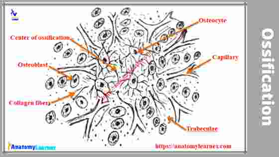
#1. You know the mesenchymal cells (osteoprogenitor cells) are the stellate shaped whose processes contact with one another. Capillaries invade in the ossification environments, and the cells become spherical, and the cytoplasmic processes become thicken.
#2. These osteoprogenitor cells differentiated into the bone forming cells – the osteoblasts.
Osteoblasts and matrix formation
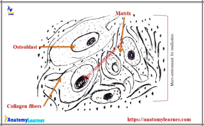
#3. These osteoblasts cells begin to synthesize and secrete osteoid and contribute to the mineralization process.
#4. First, the osteoblast cells produced the collagen fibers and appeared between the cells.
#5. In the later stage, the ground substances of the matrix are produced by the osteoblasts cells. (Osteoblasts cells produce the organic intercellular substances like mucoprotein, glycoprotein, mucopolysaccharide, and others.
#6. In early intramembranous osteogenesis, the matrix become mineralized, and osteoblasts become surrounded by this incompletely mineralized matrix. In this matrix, you might find the collagen fibers.
#7. More osteoid is produced, followed by the mineralization.
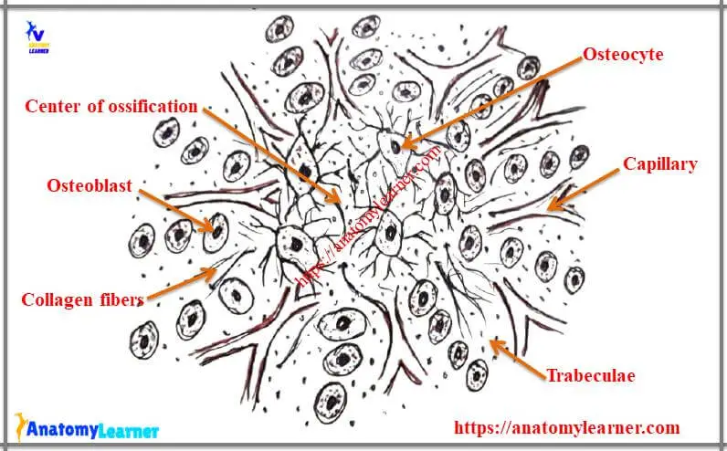
Primary ossification center formation
#8. The osteoblasts become entrapped by the surrounding matrix and transformed into osteocytes.
#9. There are small, isolated pieces of developing bones found within the mesenchyme known as the primary ossification center.
#10. Osteoblasts proliferated by division and arranged in a radiating manner from the ossification center.
#11. These bony spicules are joining with other bony spicules and form the beam or trabeculae (these are the trabecular or spongy or cancellous bone).
Mineralization process
In both intramembranous and endochondral ossification, you will find two different types of bones – immature and mature bone. Immature bone is the first bone with more bone cells and collagen fibers with less mineralized substances than mature bone.
Immature bone is also known as woven bone as there are thick bundles of collagen fibrils arranged in an irregular interanastomosing fashion. You might easily find these interanastomosing irregular collagen fibers in the woven or immature bone.
This immature bone is resorbed by osteoclasts and replaced by the mature bone. Osteoblasts synthesized and secreted organic matrix, matrix vesicle that contains lipid, alkaline phosphate, and calcium ions. All these are required for the process of mineralization.
Though mineralization is a long process but the main theme is – the alkaline phosphate produced by osteoblasts hydrolyses the locally available hexoses phosphate. Thus it produced the phosphate ions combined with calcium and become precipitated as calcium phosphate crystal in the matrix.
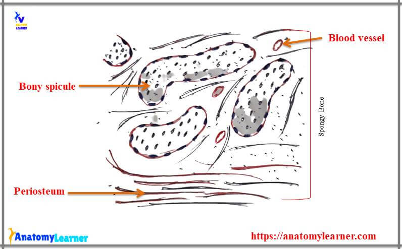
Trabecular bone to compact bone formation
The condensation of mesenchyme forms the periosteum. The vascular mesenchyme around the newly formed spongy bone condenses to form the periosteum on the outer surface and endosteum on the inner surface.
Osteogenic cells in the periosteum and endosteum differentiate into osteoblasts and start laying down the bone matrix in the form of the parallel lamella. Again, the boney trabeculae increase in width and length by adding new lamellae, and the osseous tissue replaces all the mesenchyme.
After the complete replacement of the mesenchyme, the trabecular bone becomes the compact bone. You will find the three major systems of bony lamellae arranged in an orderly pattern in the compact bone. You will find the circumferential system (outer and inner system), Haversian system, osteon, and interstitial system.
I have a complete guide on compact bone histology here in anatomy learner. If you have any interest in knowing the histology of compact bone then, you may read this article.
Intramembranous ossification vs endochondral ossification
Do you want to make a difference between the two types of ossification? Well, for this, you might know the process of endochondral ossification. Endochondral ossification is another type of bone forming environment where the bone replaces the calcified cartilage.
In intramembranous ossification, bone is formed from the condensed mesenchyme. It is a membrane model that occurs in the most flat bone of the skull and also in the clavical. You will find the primary ossification center in the whole process of this ossification.
In endochondral ossification, bone is formed from the cartilage, and it is a cartilaginous model. Most of the bones of animal’s body ossified in this process (mainly the long bone of the bone, but not the clavicle). You will find the primary center of ossification as well as the secondary center of ossification in this process.
Frequently asked question on intramembranous osteogenesis.
What is intramembranous ossification?
This is a type of microenvironment in which the bone is forming from the mesenchyme directly.
Where does intramembranous ossification occur?
It occurs mainly in the skull bone (cranial bone), face bone, and clavicle bone of animals.
What bones are formed by intramembranous ossification?
First, the spongy or trabecular bone or cancellous bone forms by this process. But after complete mineralization, it will convert into the compact bone.
Which cells contribute to the process of calcification during intramembranous ossifications?
Osteoblasts contribute to the process of calcification during this ossification process.
What type of tissue is replaced by the bone in this ossification process?
Mesenchyme tissue is replaced by the bone here in this ossification process.
“I will add other frequently asked questions on demand. You may also contribute to this FAQ section. If you have any definite questions on this type of ossification, please let me know. I will try to reply to your questions and add them here in the FAQ section.”
Labeled diagram and videos of intramembranous ossification
I already provide the video where I discussed the process of intramembranous ossification. If you found any mistakes in this video (ossification process), please let me know.
The labeled images might help you to understand the full process of ossification. If you need more images related to this ossification, you may follow anatomy learners on social media (get more updated images and notifications on updated articles).
Love to read another article from anatomy learner? Fine, you might read the following article from anatomy learner (if you want) –
#1. Spongy bone histology with identification points
#2. Understand the loose connective tissue characteristics from connective tissue histology slides
#3. Histology of dense connective tissue (dense regular and dense irregular connective tissue with slides).
Or, if you want, you may visit the histology learning laboratory, where you may start your histology learning system-wise. In most of the article, you will find real slide images and videos that will help you learn basic animal histology.
Conclusion
This long guide might help you understand the whole process of intramembranous ossification. If you think this guide is helpful, share it with your friends who want to learn the intramembranous ossification process with labeled diagrams and videos.
Please make more differences between these two types of ossification in animal’s bones. For this, you may read the other ossification guide on endochondral ossification.
If you have any inquiries about this ossification, please ask me in the comment box or direct messages. I hope you will contribute your positive thinking to enrich this guide.
Don’t forget to check my newly published articles (both in animal anatomy, histology, embryology, and comparative anatomy) here on anatomy learner. Thank you so much for staying with anatomy learner and learning anatomy with me.