The spleen of a horse is the largest hemolymph organ of its body. Here, I will describe the gross anatomy of the horse spleen with the labeled diagram.
Quick overview: a triangular-shaped spleen is situated chiefly in the left parachondric region of a horse, in close relation to the more significant part of the stomach. It presents two surfaces, two borders, and two extremities for description. The visceral surface of the horse spleen divides into two unequal parts by ridge and presents various impressions.
You will know the unique features of the equine spleen’s various surfaces, borders, and extremities. Thus, you may easily differentiate this spleen from the spleen of an ox or dog.
Horse spleen anatomy
First, I would like to enlist the unique features of the horse spleen anatomy. A horse spleen presents the below-mentioned features that make it different from other animal species –
- The spleen of a horse is roughly triangular,
- It is located at the cranial and left part of the horse’s abdomen
- between the greater part of the stomach and the diaphragm,
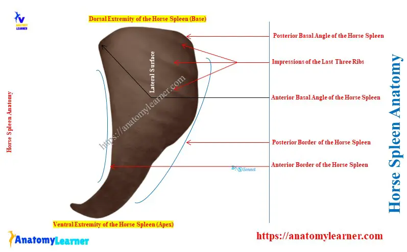
- A horse spleen presents two surfaces (parietal and visceral), two borders (anterior and posterior), and two extremities (dorsal and ventral),
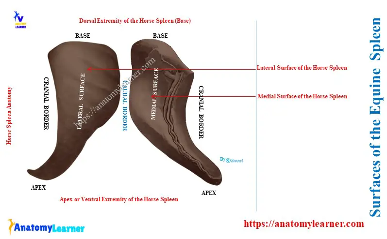
- The parietal surface is convex, and the visceral surface is divided into two unequal parts by the longitudinal ridge,
- The parietal surface of the equine spleen presents two or more impressions of the ribs,
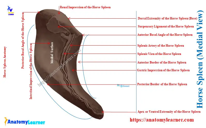
- A concave visceral surface of the equine spleen presents renal, gastric, and intestinal impressions,
- A small hilus is located at a groove in the ridge near the base of the spleen,
- The broad proximal part of the equine spleen is the base, and the lower narrow and pointed end is the apex,
- The suspensory ligament and a gastro-splenic omentum attach to the equine spleen,
Here, the diagram shows the parietal and visceral surfaces of the horse spleen with other anatomical features. Now, let’s know the details of these features from the equine spleen.
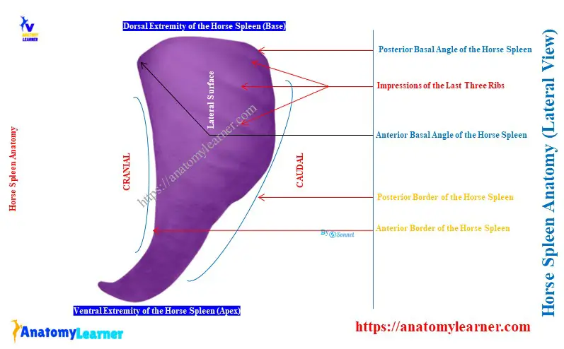
Where is the spleen located in a horse?
The spleen of a horse is located mainly in the left parachondric region. It is closely related to the left part of the greater curvature of the horse’s stomach.
This spleen extends obliquely in a curved direction between the left part of the stomach and the curs of the diaphragm. The ventral part of the spleen extends to the ventral third of the tenth or eleventh ribs of horses.
Here, the diagram shows the location and extension of the equine spleen in the parachondric region.
What is a horse spleen?
The horse spleen is a triangular organ whose size and weight vary depending on the horse’s age, breed, and health status.
The average weight of the equine spleen is about one to eight pounds. Again, the average length is about 20 inches, and the average width is about 8 to 10 inches.
In fresh condition, the equine spleen is bluish-red or purple. It is very soft and yielding in a natural state, but not friable.
Summary of the equine spleen anatomy
Here, Table 1 shows the summary of the equine spleen anatomy –
| Horse spleen | Typical features |
| Shape of the spleen | Roughly triangular |
| The parietal or lateral surface | Convex Lies against diaphragm Have contact with the last two ribs |
| Visceral or medial surface | Concave Present two unequal parts Possess renal, gastric, and intestinal impressions Present small hilus near the base |
| Anterior border | Thin and concave |
| Posterior border | Thin and convex |
| Dorsal extremity | Broad and known as base Attaches with diaphragm and sublumbar muscle |
| Ventral extremity | Narrow, pointed, and termed apex |
| Attachment of peritoneal folds | Suspensory ligament and gastro-splenic omentum |
Surfaces of the Horse Spleen Anatomy
The diagram shows the convex parietal surface of the equine spleen. Another name for this surface is the lateral surface of the spleen.
This surface chiefly lies against the horse’s diaphragm. However, it also contacts the dorsal part of the last two ribs. Sometimes, it may also extend to the flank from the lumbo-costal angle.
The visceral surface of the equine spleen, also known as the medial surface, is typically concave. A longitudinal ridge divides this surface into two unequal parts.
On this groove, you will see the hilus proximally, where the nerves and vessels are located. At the cranial aspect of the longitudinal groove, the part of the greater curvature of the stomach is attached.
Thus, the stomach forms the impression at the cranial apscet of the longitudinal groove (gastric impression), which is about one to two inches wide.
The area behind the longitudinal groove is very extensive. This area is related to the small colon, left part of the great colon, small intestine, and great omentum.
All these organs mentioned above (intestine) form the impression (known as the intestinal impression). However, there needs to be clear boundaries among these specific organs.
Borders of the equine spleen
The diagram shows two distinct borders (anterior and posterior) of the equine spleen. Here, the anterior border is thin and concave. This border is formed between the diaphragm and the greater curvature of the stomach.
Again, the posterior border of the equine spleen is also thin but convex. This border relates to the various parts of the horse intestine.
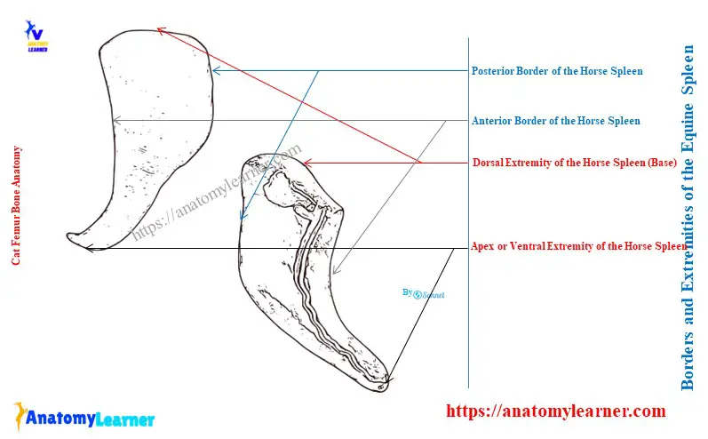
Extremities of the horse spleen
The dorsal extremity or base of the horse spleen is broad and beveled. Proximally, it fits into the interval between the left crus of the diaphragm and sublumbar muscles.
Again, it fits into the interval between the saccus caecus of the stomach and the left kidney distally. Thus, the renal impression is on the dorsal extremity of the horse spleen.
The anterior basal angle of the spleen is located at the level of the seventeenth thoracic vertebra. Again, the posterior basal angle lies against the upper part of the left flank, just caudal to the last rib.
The ventral extremity or apex of the horse spleen is small, narrow, pointed, and varies in location. Most commonly, it is opposite the tenth or eleventh rib.
Thus, the ventral extremity or apex is found just above the costal arch. Sometimes, the apex may be found further forward to the coastal arch.
Peritoneal folds of the horse spleen
The two peritoneal folds attach the equine spleen –
- A suspensory ligament, and
- A gastro-splenic omentum,
Here, the suspensory ligament of the equine spleen attaches to the dorsal end of the diaphragm’s left crus and kidney. This suspensory ligament of the spleen possesses two parts –
- Dorsal layer or part: passes to the diaphragm (ligamentum phrenico-lienale) and bends with the gastro-pherinic ligament and
- Ventral part: goes to the left kidney and is termed as the ligamentum renolienale,
Again, the gastrosplenic omentum passes from the left part of the greater curvature of the horse’s stomach. It is narrow dorsally and joins with the suspensory ligament.
Finally, the gastrosplenic omentum becomes narrow ventrally and continuous with the greater omentum.
Structure of the horse spleen
You might know the details of the histological structure of the animal spleen. The outer covering of the spleen is made of a fibrous capsule. A serous coat completely covers the fibrous capsule of the equine spleen.
Histologically, the horse spleen capsule is rich in elastic fibers and unstriped muscle tissue. The internal structure of the equine spleen includes various trabecules.
These trabeculae ramify the spleen materials to form a supporting network. Within this network, you will find dark red, soft, and grumous materials called the splenic pulp.
It contains numerous splenic and other cells and is supported by the delicate reticulum. Various branches of the splenic artery enter into the hilus and pass along the trabeculae.
You will also find the splenic lymph node in the equine spleen structure.
Vessels and nerves to the equine spleen
The splenic artery supplies the equine spleen. It is a branch of the horse celeic artery. Various branches of the splenic arteries supply the equine spleen.
The horse splenic vein lies caudal to the splenic artery in the hilus. This splenic vein goes to the portal vein.
The sympathetic celiac plexus nerve innervates the horse’s spleen.
Conclusion
So, the horse spleen anatomy consists of two surfaces, two borders, and two extremities. It extends obliquely in the curved direction in the parachondric region of the horse.