The horse pancreas is a triangular soft gland in its digestive system. This gland of the horse has both exocrine and endocrine functions. Here, I will describe the anatomy of the horse pancreas with the labeled diagram.
Quick overview: The horse pancreas is triangular in outline and presents two surfaces, three borders, and three angles for description. It invariably possesses two ducts—one pancreatic duct and another accessory pancreatic duct.
You will know the exact location and relationship of the equine pancreas with other organs. I will describe every surface, border, and angle of the pancreas with the diagram.
Again, I will also show the ducts that arise from the equine pancreas with their site of opening. So, let’s get started to learn the details of the anatomical features of the horse pancreas.
Where is the pancreas located in a horse?
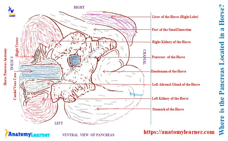
The horse pancreas is located transversely on the dorsal wall of the abdomen. Its most significant part is the right of the median plane.
The central part of the equine pancreas lies under the sixteenth and seventeenth thoracic vertebrae. This gland is also related to the kidneys, adrenal bodies, spleen, vena cava, caecum, and colon.
Let’s see the relationship of the equine pancreas with the different organs of the abdomen –
- The horse pancreas is dorsally attached to the kidney and adrenal bodies with connective tissue. It also attaches to the gastro-phrenic ligament, posterior vena cava, portal fissure, and suspensory ligament of the spleen.
- Ventrally, the pancreas is attached to the base of the caecum and the end part of the colon by the areolar tissue.
Here, the diagram shows the relative position of the equine pancreas in the abdominal cavity.
Appearance and color of the equine pancreas
Typically, the equine pancreas is a triangular-shaped organ of the abdomen. But, when it preserves and hardens, its shape becomes very irregular.
The external appearance resembles the salivary gland. But this pancreas is soft and possesses loosely arranged lobules.
In fresh condition, it has a reddish cream color. It gradually becomes dark if it is left for an unpreserved sample.
The average weight of the equine pancreas is about 350 – 400 grams.
Horse pancreas anatomy
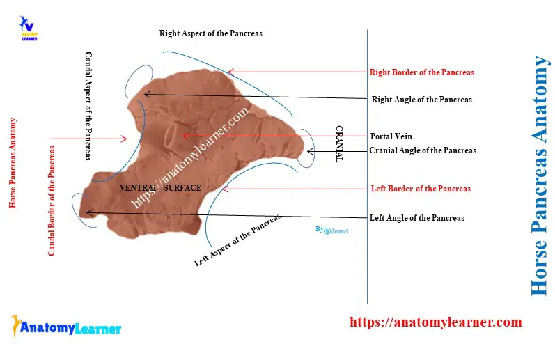
You might describe the features of the various surfaces, borders, and angles in the horse pancreas anatomy. Let’s see the below-mentioned surfaces, borders, and angles from the equine pancreas –
- Dorsal surface: faces dorsally and forward,
- Ventral surface: faces ventrally and backward,
- Right border: it is nearly straight,
- Left border: this border is slightly concave,
- Posterior border: this border of the equine pancreas presents a deep notch,
- Cranial or duodenal angle: this is the most ventral part of the pancreas,
- Left or splenic angle: it is the cranial irregular part of the pancreas,
- Right angle of the pancreas: this angle is rounded,
- Ducts of the pancreas: consists of pancreatic and accessory pancreatic ducts,
The horse pancreas labelled diagram identifies the surfaces, borders, angles, and ducts. Now, let’s know the details of the various surfaces, borders, and angles of the pancreas.
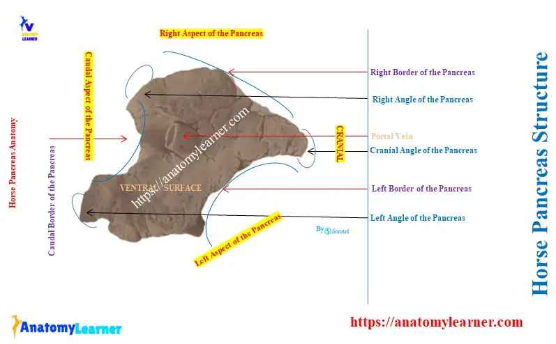
Surfaces of the equine pancreas
The peritoneum partially covers the dorsal surface of the horse’s pancreas. This surface of the pancreas is chiefly related to the following organs –
- The ventral surface of the right kidney and adrenal gland,
- Caudal vena cava, portal vein, and celiac artery,
- The gastro-phrenic ligament and the caucus caecus of the horse stomach,
- Right and caudate lobes of the horse liver, and
- Gastro-pancreatic folds,
You will see several grooves on the dorsal surface of the horse pancreas anatomy. Some grooves are for the divisions of the celiac artery. The larger groove on the dorsal surface of the equine pancreas is for the splenic vein.
The ventral surface of the equine pancreas is generally concave. It has two impressions, separated by an oblique ridge.
The smaller impression present on the ventral surface lies to the right. It is caused by the pressure of the base of the caecum.
Again, the larger impression is located in the area of the terminal part of the greater colon. The peritoneal covering is not visible on the ventral surface of the equine pancreas, but a small area at the cranial angle shows it.
Borders of the horse pancreas
Here, the horse pancreas’s right, left, and caudal borders are identified. The right border is related to the second part of the horse’s duodenum.
The concave left border is related to the first part of the duodenum, the left sac of the stomach, and the splenic vessels.
The root of the great mesentery is in contact with the deep notch on the caudal border of the equine pancreas. The portal vein, which passes through the pancreas obliquely, lies on the right side of the notch.
A thin bridge of gland tissue dorsal to the portal vein helps form the portal ring in the horse.
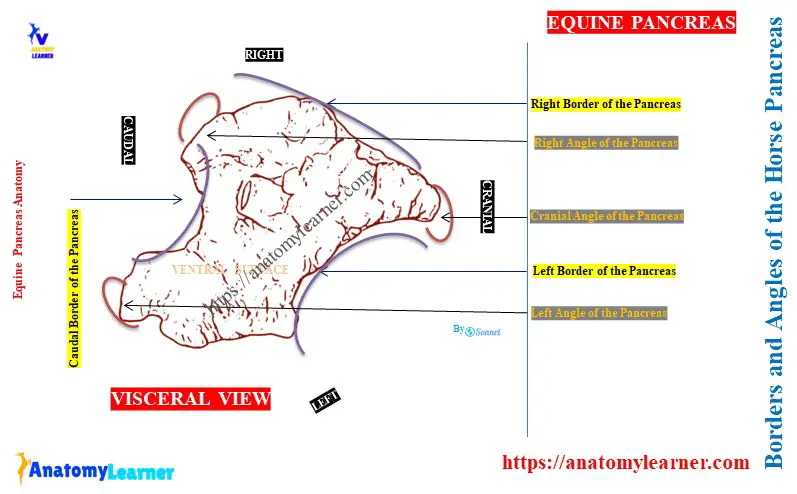
Angles of the horse pancreas
The anterior or duodenal angle attaches to the concavity of the second curve of the duodenum. It also attaches to the adjacent part of the right lobe of the liver. The pancreatic ducts leave the pancreas from this extremity.
The left or splenic angle of the horse pancreas fits into the space between the saccus caecus of the stomach in front. It also relates to the left kidney caudally, the base of the spleen dorsally, and the terminal part of the great colon ventrally.
The right angle of the pancreas lies on the ventral surface of the right kidney and adrenal gland.
Pancreatic ducts of the equine pancreas
Two variable pancreatic ducts exist in the horse pancreas. The larger one is the main pancreatic duct, whereas the smaller one is termed the accessory pancreatic duct.
The pancreatic duct of a horse is one centimetre wide and has a thin wall. It is located in the pancreas near its dorsal surface.
This pancreatic duct is formed by the union of two radicles from the right and left extremities. It passes through the duodenal angle and opens at the duodenal diverticulum.
On the other hand, the accessory pancreatic duct of the horse arises from the pancreatic duct. It opens on a papilla in the duodenum opposite to the opening of the horse’s pancreatic duct.
Vessels and nerves innervation
The arteries that supply the horse pancreas come from the branches of the celiac and cranial mesentery arteries. Again, the veins that supply the pancreas go to the portal vein.
The sympathetic plexuses from the celiac and mesenteric nerves innervate the horse pancreas.
Species differences in the pancreas
The small ruminant and cat possess only a single pancreatic duct. Again, the pancreas of the ox, pig, and dog possesses the accessory pancreatic duct.
Both pancreatic and accessory pancreatic ducts are present in the pancreas of horses and dogs, and they are occasionally present in the ox’s pancreas.
The pancreatic duct is smaller than the two accessory pancreatic ducts in the dog pancreas.
Conclusion
So, the roughly triangular-shaped pancreas is located transversely on the dorsal wall of the horse’s abdomen. The lobules are loosely unit in the horse pancreas anatomy and present two surfaces, three borders, and three angles.