The horse esophagus anatomy divides into cervical, thoracic, and small abdominal parts. It is a Musculo-membranous tube in horses that extends from the pharynx to the stomach.
Here, I will describe the course, parts, and relationship of the horse esophagus with other organs with the diagram.
Quick overview of the equine esophagus: it is a Musculo-membranous tube that extends from the pharynx to the stomach and divides into three parts. The esophagus’s diameter gradually increases and relates to various organs of the cervical and thoracic regions.
This guide also explains the main differences among the esophagus of dogs, cows, and horses. Let’s start by learning about the horse esophagus’s features and courses.
Horse esophagus anatomy
First, let’s see the unique features of the horse esophagus anatomy –
- It is a tube-like Musculo-membranous structure,
- It extends from the pharynx to the stomach,
- The equine esophagus divides into three major parts – cervical, thoracic, and abdominal parts,
- At the origin, the esophagus relates to the cricoid cartilage and cricoid-arytenoid muscle,
- It ends at the cardiac orifice of the stomach,
- Structurally, the esophagus consists of four layers – mucosa, submucosa, muscular coat, and adventitia,
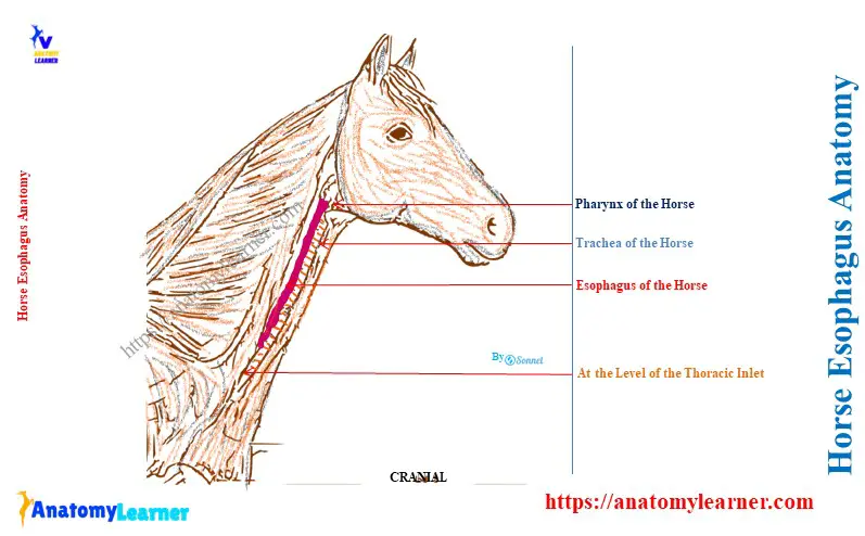
Here, the diagram shows the course overview and parts of the equine esophagus. You will also find various diagrams of the equine esophagus throughout this guide.
What is a horse esophagus?
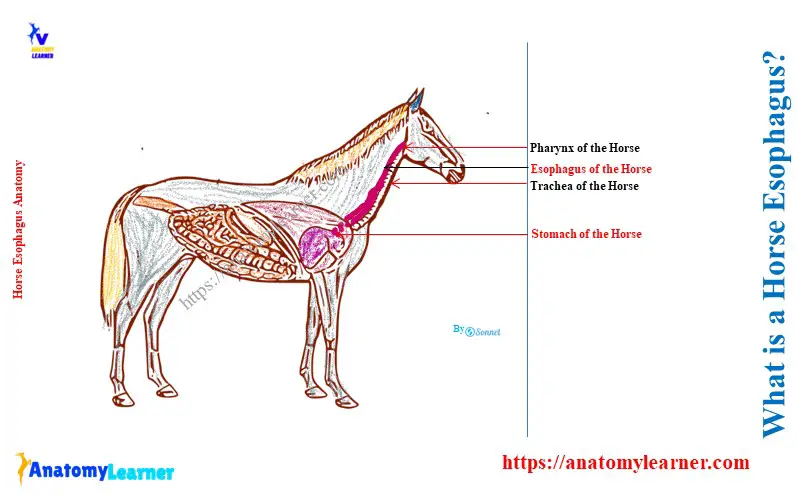
The horse esophagus is the first part of the alimentary tract of its digestive system. It is a typical tubular structure that transports food from the oral cavity and pharynx down the neck and through the thorax to the stomach.
An extra abdominal part is present in the horse esophagus compared to the ox esophagus. A keratinized stratified squamous epithelium lines and protects the equine esophagus.
Parts of the equine esophagus
If you observe the equine esophagus in the horizontal plane, it courses downward and backward. It continues in this direction until it enters the thorax and passes upward to reach the dorsal face of the trachea.
After a short distance, the direction of the esophagus becomes almost horizontal. Then, the equine esophagus passes somewhat upward and terminates at the stomach.
You will find the detailed course of this equine esophagus in the next section of this guide. During its courses, you will see three parts according to the region of the horse’s body –
- A large cervical part (Pars cervicalis): it is the largest part of the esophagus among three parts,
- The thoracic part (Pars thoracalis): it is shorter than the cervical part of the esophagus and
- A small abdominal part (Pars abdominalis) is the smallest part of the esophagus in the equine. However, you will not find any abdominal part in the cow esophagus due to the close apposition of the stomach and diaphragm.
How long is a horse’s esophagus?
The average length of a horse’s esophagus is about 125 – 140 centimeters. Here, the cervical part of the esophagus is about 65 – 75 centimeters long.
The average length of the thoracic part of the equine esophagus is about 57 – 60 centimeters. Finally, the small abdominal part of the esophagus is about 3 – 5 centimeters.
The course of the Horse Esophagus Anatomy
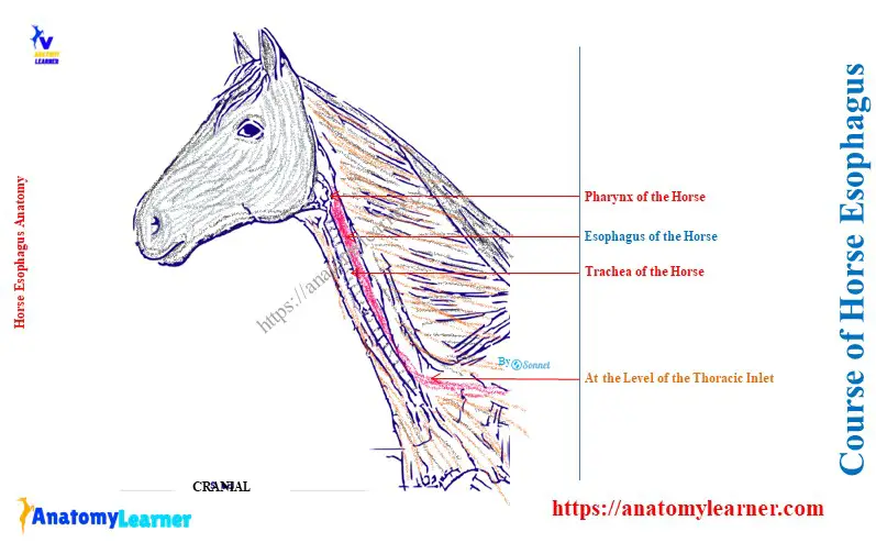
The horse esophagus shows several changes in the direction in its courses. Let’s see the course of the esophagus from the larynx to the stomach –
- The esophagus starts at the level of the anterior border of the cricoid cartilage of the pharynx. In this situation, it remains in the median or longitudinal plane of the body.
- At the fourth cervical vertebra level, the esophagus goes to the left side of the trachea. This relationship continues with the trachea up to the third thoracic vertebra.
- Now, the esophagus goes to the dorsal surface of the trachea again. It passes backward and crosses the aortic arch of the aorta. Thus, it pushes over to the right of the median plane.
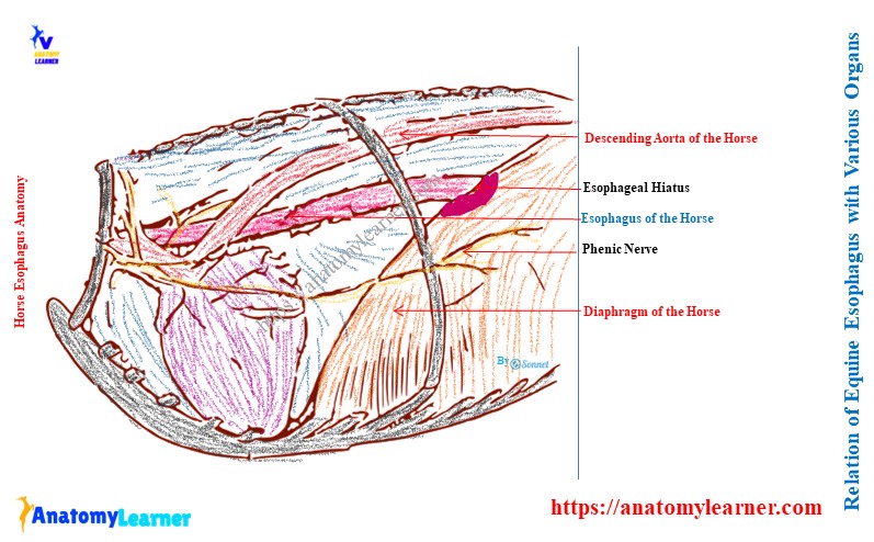
- Now, the esophagus continues to the mediastinum space between two lungs and goes backward a little dorsally. Gradually, it goes to the left side of the median plane and reaches the esophageal hiatus of the horse’s diaphragm.
- After passing through the esophageal hiatus, the esophagus ends at the cardiac orifice of the horse’s stomach. The termination occurs just a few inches ventral to the vertebral extremity of the fourteenth rib.
Related article: How many ribs does a horse have?
Relation of horse esophagus with various organs
Here, I will describe the major relationships of the horse esophagus with the various organs in different regions.
At the origin of the esophagus, it is related to the following different organs –
- Dorsally: the origin of the esophagus relates to the guttural pouches and the ventral straight muscles,
- Ventrally: circoid cartilage and dorsal crico-arytenoid muscle, and
- Laterally: carotid arteries,
In the middle of the neck, the equine esophagus is related to the following organs –
- Dorsally: left longus colli muscle,
- Laterally: left carotid artery, vagus nerve, sympathetic fiber, recurrent laryngeal nerve, and
- Medially: trachea,
At the thorax, the equine esophagus is in contact with the below-mentioned organs –
- Near the thoracic inlet: left jugular vein for a short distance,
- In thorax: medial side of trachea, first rib, root of the brachial plexus, and left caudal cervical ganglion laterally,
When the esophagus remains on the dorsal to trachea, it also has contact with the aorta, vena azygos, and right vagus. Here, the aorta remains on its left aspect. Again, the vena azygos and right vagus nerve remain on the right side of the esophagus.
Structure of the equine esophagus
The wall of the equine esophagus is composed of four distinct coats –
- Inner mucous membrane coat or tunica mucosa,
- A submucosa layer or tunica submucosa,
- The muscular coat or tunica muscularis, and
- An outer fibrous layer or tunica adventitia,
You may learn the details and features of these four coats from the histology of a tubular organ.
What muscles are in the esophagus of a horse?
The esophagus of a horse contains both the striped and unstriped muscles. Here, the tunica muscularis coat of the wall of the equine esophagus is typically striped in nature. But, it changes as far as it passes caudally and reaches the base of the heart; it becomes an unstriped type of muscle.
Again, with these changes, the muscular layer also gradually becomes thicker and firmer caudally. The lumen of the esophagus also gradually decreases in horses.
Except at the end of the tube, the tunica muscularis of the esophagus consists of two layers of muscles arranged spirally. They may also arrange elliptically and intercross dorsally and ventrally.
At the origin of these two bundles of muscles, they are nearly 2 inches wide. These muscles arise from the caudal part of the pharyngeal raphe and the common tendon of the cricopharyngeal and they-pharyngeal muscles.
At the terminal part of the esophagus, its tunica muscularis layer shows an external longitudinal and an internal circular layer of muscles. Thus, the end part of the equine esophagus becomes thicker than the proximal or cranial part.
What is the mucous membrane of the horse esophagus?
The mucous membrane is a pale and internal layer of the tubular esophagus. A simple stratified squamous epithelium lines the mucous membrane of the horse esophagus.
This mucous membrane is loosely attached to the tunica muscularis layer by a thick submucosa layer. Several longitudinal folds are present on the tunica mucosa, or mucous membrane of the esophagus.
Vessels and nerves of the equine esophagus
The carotid, broncho-esophageal, and gastric arteries supply to the equine esophagus. Again, the vagus, glosso-pharyngeal, and symphathetic nerves innervate the equine esophagus.
Species differences in the esophagus
The basic difference among the esophagus of a horse, ox, and dog are given in Table 1 –
| Esophagus | Horse | Ox or cow | Dog |
| Length | Longer 125 – 140 cm | Shorter than horse 85 – 90 cm | Length variable |
| Parts | Two parts Cervical and thoracic | Three parts Cervical, thoracic, and abdominal | Three parts Very short abdominal part |
| Wide | Wider | Less wide | Comparatively wider |
| Constriction | N/A | N/A | Constriction at the beginning |
Conclusion
Thus, the horse esophagus anatomy is a tubular structure that extends from the pharynx to the cardiac orifice of the stomach. The course of the equine esophagus is slightly different from that of the cow esophagus.
Again, the equine’s esophagus has an extra distinct abdominal part compared to the cow’s esophagus. The end part of the horse’s esophagus becomes thicker than the cow’s esophagus.