
The horse sternum is a long plate of osteo-cartilagenous structure. Here, I will describe its anatomy in detail with the diagram.
Quick overview: the horse sternum is a boat-shaped bone formed by the fusion of seven sternebrae. Anatomically, it presents three surfaces, three borders, and two extremities.
You will learn the features of these surfaces, borders, and extremities from the equine sternum. Finally, you will easily differentiate it from the sternums of other species, such as the ox, dog, and pig.
Horse sternum anatomy
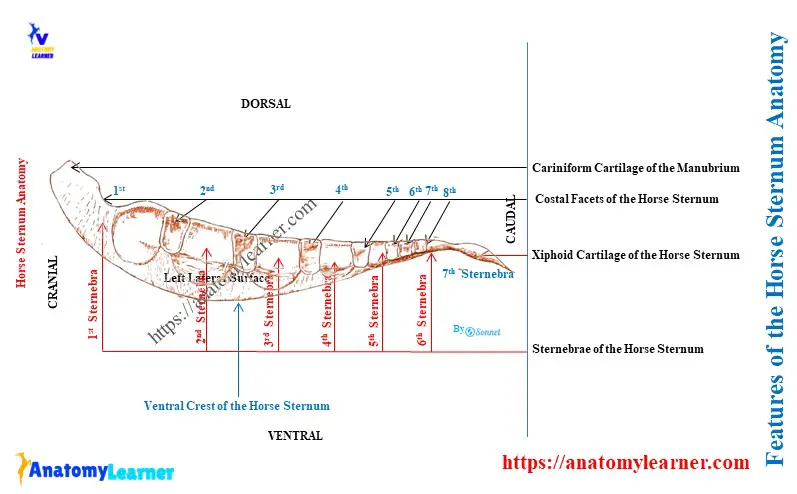
Here, the diagram presents the features of the horse’s sternum anatomy. Let’s try to identify the below-mentioned structures from the real equine sternum –
- Three surfaces: one dorsal and two lateral,
- Three borders: two dorsolateral and one ventral border,
- Two extremities: cranial manubrium and caudal xiphoid cartilage,
- Seven segments from the sternum,
- Seven pairs of costal cavities or facets on the dorsal surfaces and
- Ventral crest on the ventral border of the sternum,
Now, let’s see the unique features of the horse sternum –
- The sternum is a boat or canoe shape,
- It is formed by the fusion of seven sternebrae (segments of the sternum),
- The equine sternum is compressed laterally towards the cranial end and dorsoventrally at the caudal end,
- The first segment is the manubrium, which possesses a cranial cartilaginous extension,
- The last segment is the xiphoid cartilage, which is flat and rounded,
- Two lateral surfaces (right and left) are convex and possess seven costal facets (on each side), and
- The ventral border of the sternum possesses a prominent ventral crest that fades out caudally,
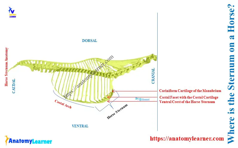
Where is the sternum on a horse?
A horse’s sternum is placed at the midline of the floor of the thoracic cavity. It is compressed laterally, except in its caudal part, which is flattened dorsoventrally.
The equine sternum inclines obliquely so that the caudal end is about 6 – 8 inches lower than the cranial. The costal cartilage of the first eight-pair ribs attaches to the corresponding costal facets of the equine sternum.
How many sternebrae are there in a horse?
There are 7 sternebrae in the horse sternum, from the first to the seventh. The first and last sternebrae are highly modified in horses compared to the ox sternebrae.
The number of the sternebrae of the sternum is different in various animals –
- The dog has eight sternebrae in its sternum,
- A pig has six sternebrae in its sternum,
- But, the rabbit and ox possess seven sternebrae in their sternum,
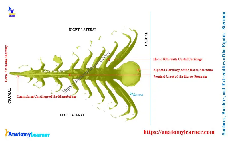
Surfaces of the horse sternum anatomy
The surfaces of the horse sternum are different from those of the ox sternum. The ox sternum has dorsal and ventral surfaces, whereas the horse sternum possesses one dorsal and two lateral surfaces. The ventral surface is practically absent from the equine sternum.
The dorsal surface of the equine sternum is a very narrow isosceles triangle shape with a cranial apex. This surface is concave longitudinally and flattened transversely.
Both the right and left lateral surfaces are convex above. They are slightly concave below and diminish in extent behind.
Each dorsolateral aspect of the lateral surfaces contains seven costal cavities. The sternal end of the second to eight costal cartilage makes inclusive articulation with the corresponding costal facets.
All these coastal facets are located in series at the intervertebral junctions. The first four costal facets are elliptical in outline and have a long vertical diameter. They are separated by a considerable regular interval.
The rest of the costal facets of the equine sternum are progressively smaller. They are more circular and closer together. The pectoral muscles attach below the area of these costal facets.
Borders of the horse sternum
Practically, the horse sternum has one ventral and two dorsolateral borders. Here, the dorsal and lateral surfaces meet dorsally and form the dorsolateral surface on the right and left sides.
The dorsolateral borders of the sternum give attachment to the lateral branches of the sternal ligament. However, the lateral border of the ox sternum presents seven pairs of sternal facets on either side of the intersternal junctions.
Two lateral surfaces meet ventrally and form the ventral border. Here, the ventral border of the equine sternum shows the prominent keel-like crest of the sternum.
You may easily feel this crest of the ventral border of the sternum from the horses. But the crest fades out caudally.
Extremities of the equine sternum anatomy
The manubrium sterni forms the cranial extremity of the equine sternum. You may easily feel this manubrium sterni from the live horse from its central furrow of the pectoral region.
The manubrium sterni of the horse consists of a laterally compressed large cartilaginous prolongation. This cartilaginous prolongation is known as the carniform cartilage in horse.
The lateral surface of this carniform cartilage is flat and furnishes attachment for the pectoral muscles. A round ventral border is present in the carniform cartilage, continuing caudally with the body of the sternum.
The dorsal border of the carniform cartilage is concave. It possesses an articular cavity for the first pair of costal cartilage.
The xiphoid cartilage forms the caudal extremity of the equine sternum. It is a thin plate connected infornt with the last bony segment of the sternum.
The connected part of the xiphoid cartilage is narrow and thick, termed the neck. But, the caudal and lateral part of the xiphoid cartilage is expanded and nearly rounded.
The xiphoid cartilage presents a dorsal and ventral surface. Here, the dorsal surface of the xiphoid cartilage is concave and provides attachment to the diaphragm.
Again, the ventral surface of this cartilage is convex. It provides attachment to the transverse abdominis muscles and the linea alba. The free margin of the horse’s xiphoid cartilage is very thin.
Development of equine sternum
At the birth, the horse sternum anatomy consists of 7 sternebrae or bony segments. These seven sternebrae join together by the intersternal cartilages.
The last two sternebrae fuse in the second month of the birth. However, the other sternebrae do not usually unite completely, even in older age.
All the sternebrae consist of a very vascular spongy bone. This spongy bone of the sternebrae is covered with a very thin layer of a compact substance.
Thus, the equine sternum consists to a considerable extent of persisting cartilages. In older age, they may undergo partial ossification.
Species differences in the sternum
Table 1 shows the significant differences among the sternum of the horses, dogs, and cows –
| Species | Cow Sternum | Horse Sternum | Dog Sternum |
| Shape | Wide, flatter, and longer Compressed dorsoventrally | Canoe shape Compressed laterally except the caudal end The caudal end is flattened dorsoventrally | Long, laterally compressed |
| Number of the segments | 7 sternebrae in cow 6 sternebrae in goat | 7 sternebrae | 8 sternebrae |
Conclusion
The sternum of a horse anatomy is shaped somewhat like a canoe. It is compressed laterally except in its caudal part. The anatomy of a horse’s sternum consists of 3 surfaces, three borders, and two extremities for description.