
The horse’s liver is the largest gland in its body. Here, I will describe the anatomy of the horse liver in detail with the diagram.
Quick overview: the horse liver is thin, and extensive glands are placed obliquely on the abdominal surface of the diaphragm. Anatomically, it possesses two surfaces, four borders, and three main lobes.
All the features from the surfaces, borders, and lobes are identified from the horse liver. I will also describe the ligaments that hold the liver in its position.
Horse liver anatomy
First, let’s identify the below-mentioned features from the horse liver anatomy –
- Parietal and visceral surfaces,
- Portal fissure on the visceral surface with contents,
- Various impressions at the visceral surface: gastric, colic, duodenal, and cecal impressions,
- Borders of the liver: dorsal, ventral, right, and left borders,
- Renal impression on the dorsal border, and umbilical fissure on the ventral border,
- Three main lobes of the liver: right, middle, and left, and
- Five or six ligaments of the liver,
All these features mentioned above are identified from the labeled diagram of the equine liver. You will also find other diagrams on the equine liver that are labeled differently throughout this guide.
The horse liver is somewhat unique compared to the ox liver. Let’s see the unique features of the horse liver –
- It is very extensive and thin,
- It is placed obliquely on the abdominal surface of the diaphragm,
- The major part of the equine liver is located on the right side of the median plane.
- A fissure at the ventral border divides the liver into three main lobes – right, middle, and left,
- The right lobe of the liver possesses the caudate lobe and processes,
- Both the right and left borders of the equine liver are thin,
- The gall bladder is absent in the equine liver,
- The hepatic duct is longer and opens directly into the duodenum behind the pyloric end of the stomach,
- There is a hepato-pancreatic ampulla on the duodenum where the pancreatic duct opens with the hepatic duct,
- The number of ligaments is more in the equine liver than in the ox liver,
Let’s describe the surfaces, borders, lobes, and ligaments from the anatomy of the equine liver. Before that, let’s know the exact location, color, and size of the equine liver.
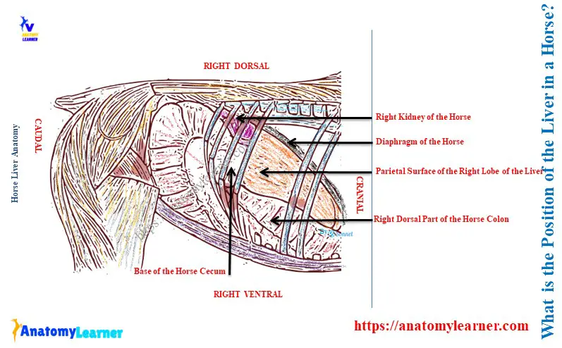
What is the position of the liver in a horse?
The position of the horse’s liver is on the right side of the abdominal cavity in an oblique downward and forward direction. Here, the upper part of the liver is at the level of the right kidney. Again, the lower part is located on the left side of the abdomen.
The lower part is located above three to four inches from the abdominal floor, opposite to the ventral end of the seventh or eighth ribs. The greater part of the horse’s liver lies right to the median plane of the body.
Horse liver size and color
The average weight of the horse’s liver is about 5 – 6 kg. But, the weight may be 8 – 10 kg in the large draft horse breed.
Within the body cavity, the liver is strongly curved and adapted to the abdominal surface of the diaphragm. But, when it is removed from the body cavity, it becomes flattened and covert into a cake-like form.
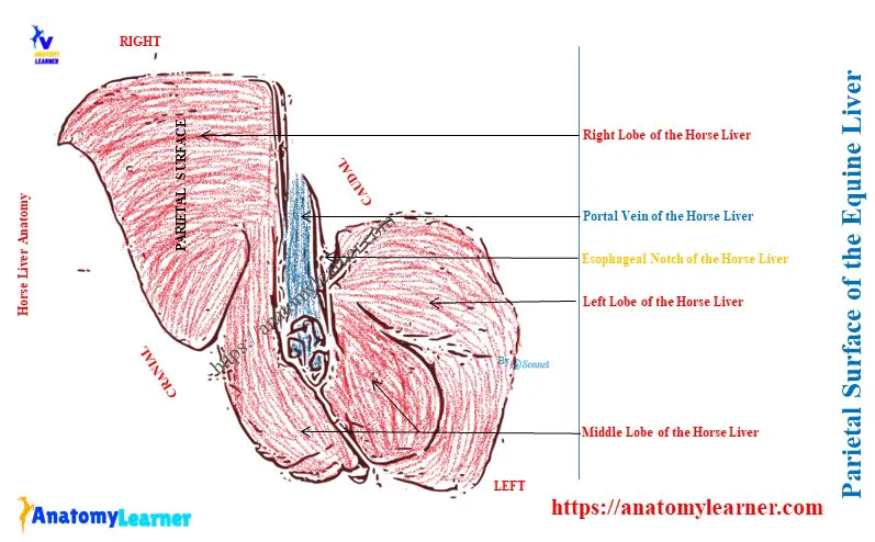
The parietal surface of the equine liver
Here, the diagram shows the parietal surface of the equine liver anatomy. The parietal surface of this liver is strongly convex and lies against the diaphragm.
The parietal surface of the liver is directed, usually dorsally and forward. The most cranial part of this surface is opposite the ventral third of the sixth intercostal space or seventh rib.
To understand the position of the parietal surface, you might know a horse’s ribs. There are a total of thirteen pairs of ribs in the horse skeleton.
The parietal surface of the liver presents a sagittal groove for the caudal vena cava. This sagittal groove is located just to the right of the median plane. Again, this groove partially embeds into the substance of the liver gland.
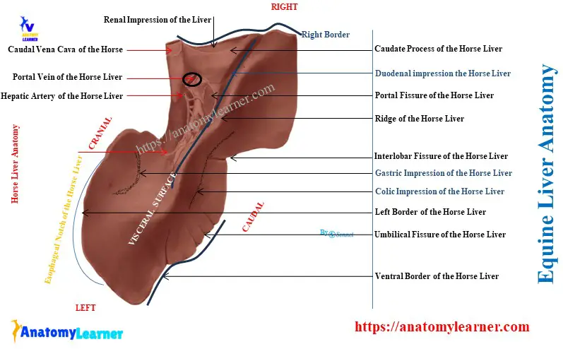
Visceral surface of the horse liver anatomy
Here, the diagram shows the features from the visceral surface of the horse liver anatomy. The visceral surface of the liver is directed ventrally and backward.
It is irregular, concave, and molded on the organ against it. You will find the following features on the visceral surface of the equine liver –
- Portal fissure on the proximal extremity,
- Extensive gastric impression,
- Longer duodenal impression,
- Larger colic impression, and
- Small caecal impression,
Let’s learn the details of these structures from the visceral surface of the liver using the labeled diagram.
Portal issue of the equine liver
It is a depression above the middle of the visceral surface of the liver. This fissure is located just a little to the right of the median plane.
Structures that enter through the portal fissure of the liver:
- The portal vein of the horse,
- Hepatic artery, and
- Hepatic plexus of the nerves,
The structure that leaves from the portal fissure of the liver:
- Hepatic duct and
- Lymph vessels of the horse’s liver,
You will also find the hepatic lymph glands on the portal fissure of the equine liver. The lesser omentum surrounds the portal fissure, and the pancreas attaches to its right side.
You will see the caudate lobe of the liver just above the portal fissure. This caudate lobe continues to the right by the caudate process.
Impressions of the equine liver
The gastric impression is an extensive concave area on the visceral surface of the liver. This gastric impression is the surface of the contact with the stomach. It is located below the portal fissure and above the colic impression of the equine liver.
The duodenal impression on the visceral surface is longer. It is located above the gastric impression and on the right of the median plane.
The colic impression is located ventrally and to the right of the gastric and duodenal impressions. Among these three impressions of the liver, you will see a ridge that provides an extensive contact surface for the diaphragmatic flexure and the right dorsal part of the colon.
Sometimes, a small caecal impression may be found at the dorsal to the colic impression. It provides a surface for contact with the cranial part of the base of the caecum.
Borders of the horse liver
Most areas of the dorsal border of the horse liver are thick. You will find the below-mentioned structures on the dorsal border of the equine liver –
- The attachment of the right lateral ligament,
- A depression in the right kidney of the horse (renal impression),
- A notch for the caudal vena cava,
- The deep esophageal notch and
- The attachment of the left lateral ligament,
Here, the esophageal notch is partly occupied by the end of the esophagus but chiefly by the thick margin of the esophageal hiatus.
The ventral border of the liver is thin and marked by two interlobar fissures. These fissures partly divide the liver into the right, middle, and left lobes.
The right lobe of the liver is larger, whereas the middle one is smallest. The middle lobe is marked by various small fissures and umbilical fissures. It contains the umbilical vein in the fetus, which converts into the round ligament after birth.
The right border of the equine liver is thin and long. It extends backward mainly to the sixteenth rib, a little below its middle.
Finally, the left border of the liver is also thin and convex. This border starts at the left side of the esophageal notch, about a handbreadth ventral to the fourteenth thoracic vertebra.
It curves downward, outward, and forward to a point opposite to the ventral end of the ninth rib. Now, it runs forward about parallel to the costal arch as far as the ventral end of the seventh rib.
Here, the ventral and lateral borders of the equine liver form the margo acutus.
What are the lobes of the liver of a horse?
The right, middle, and left are the three principal lobes of a horse’s liver. Here, the right lobe of the young horse is the largest and irregular quadrilateral form.
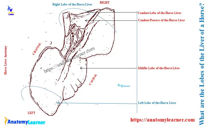
The caudate lobe is located on the dorsal part of the right lobe. The caudate lobe ends in a pointed caudate process that is directed outward. It forms the renal impression for the right kidney with the right lobe.
The middle lobe of the horse liver is comparatively smaller compared to other lobes. Another name for the middle lobe is the central lobe.
The left lobe of the equine liver is oval and thickens centrally. Middle and right lobes become smaller in most of the middle-aged horses.
How is the hepatic duct formed in the equine liver?
The hepatic duct is formed by the union of the right and left chief lobar ducts. It occurs at the portal fissure of the equine liver.
This hepatic duct is about 5 centimeters long and 1 – 1.5 centimeters wide in a horse. It passes between two layers of the mesoduodenum. The hepatic duct now pierces the duodenal wall just 12 – 15 centimeters behind the pylorus, along with the pancreatic duct.
This duct obliquely passes through the duodenal wall before opening into the diverticulum duodeni.
Ligaments of the horse liver anatomy
The liver of the horse is held in position largely by –
- The pressure of the other viscera, and
- By its close application and attachment to the diaphragm,
You will find the below-mentioned ligaments in the horse liver anatomy –
- A coronary ligament,
- The falciform ligament,
- A round ligament,
- The right lateral ligament,
- The left lateral ligament and
- A hepato-renal or caudate ligament,
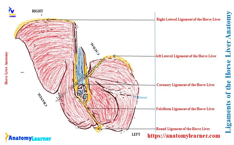
Coronary ligament of the equine liver
The coronary ligament of the equine liver closely attaches it to the diaphragm. It consists of two strong laminae – right and left.
Here, the right lamina attaches to the right fossa of the vena cava. Again, the left lamina begins on the left of the vena cava and passes dorsolaterally.
It continues with the left lateral ligament at the left margin of the esophageal notch of the liver. Again, it detaches a middle fold that extends to the esophageal notch and continues with the lesser omentum.
Falciform ligament of the liver
The right and left laminae of the coronary ligament unite below the vena cava and form the falciform ligament. It is a crescentic fold-like structure in the liver. It attaches to the middle lobe, the sternal part of the diaphragm, and the abdominal floor.
Round ligament of the equine liver
The round ligament is the fibrous cord-like structure on the concave edge of the falciform ligament. It extends from the umbilical fissure to the umbilicus.
And you know, the round ligament is the vestige of the fetus’s umbilical vein. In the fetus, it supplies to the liver from the placenta.
Right lateral ligament of the liver
The right lateral ligament of the liver is long and strong. It attaches to the dorsal border of the right lobe closely to the costal part of the diaphragm.
Left lateral ligament of the liver
The left lateral ligament is an extra ligament in the horse liver compared to the ox liver. It is a triangular fold-like structure in the equine liver.
This left lateral ligament of the equine liver attaches to the dorsal edge of the left lobe to the tendinous center of the diaphragm.
Hepatorenal or caudate ligament of the equine liver
This ligament attaches to the caudate process to the right kidney and base of the caecum. Here, the lesser omentum and the first part of the mesoduodenum are formed by the peritoneum, leaving the visceral surface at the portal fissure.
It also leaves from the fissure to the esophageal notch of the liver. Finally, they pass to the lesser curvature of the horse’s stomach and the first part of the duodenum.
Horse liver structure
An external serous and internal fibrous coat covers the horse’s liver. Here, the serous coat covers the liver except at the attachment of the pancreas and at the portal fissure. It contributes to the ligaments and lesser omentum.
The fibrous coat of the liver is comparatively thin and sends laminae into the ligaments. It also sends the trabeculae into the stroma of the liver tissue.
The fibrous coat is abundant at the portal fissure of the equine liver. This coat also surrounds the vessels and ducts of the equine liver.
The liver substance consists of parenchyma and stroma. Here, the parenchyma consists of polygonal lobules held together by a small amount of interlobular connective tissue.
Again, the horse liver lobule consists of polyhedral liver cells, delicate fibers, capillaries, and a central vein.
Conclusion
So, the horse liver anatomy presents two surfaces and a circumference that may divide into four borders. It also presents three major lobes and six distinct ligaments.