
Dense connective tissue is white that has less flexibility and far more resistant to stress. You know, dense connective tissue divides into dense regular connective tissue and dense irregular connective tissue based on fibers orientation.
Hey, if you want to learn the basic histology of dense connective tissue, you might know the histology of dense irregular connective tissue. This short post will discuss the histology of dense irregular connective tissue with slide images under the light microscope.
You will learn the fibers’ orientation (especially the collagen and elastic fibers) and cells from the dense irregular tissue. You will get the identification points that might help you identify the histology slide of dense irregular tissue samples under a light microscope.
Fine, let’s get into the main parts of today’s post–dense irregular connective tissue histology with slide images and diagrams under the light microscope.
Dense irregular connective tissue histology
Before starting learning dense irregular connective tissue histology, you might have basic knowledge of the different types of connective tissue cells and fibers, especially the collagen and elastic fibers and ground substances. I want to enlist the important histological features from the real slide of dense irregular tissue that you might identify. Let’s have a look at these important histological structures –
- #1. Thick collagen fibers and bundles
- #2. Thin elastic fibers (branching and anastomosing)
- #3. Fibrocytes in the densely packed collagen bundles
- #4. Nuclei of fibrocytes
- #5. Blood vessels on the sample connective tissue sample
Okay, now you might find out and identify these structures from the sample tissue sample.
Identification of dense irregular tissue slide
If you want to identify the dense irregular connective tissue slide under the light microscope, you might know all the histological features (describe later) of this tissue. Here I will enlist the identification point that might help you to identify dense irregular tissue microscope slide under the light microscope.
#1. Presence of thick, densely packed collagen bundles that oriented randomly in the sample tissue section.
#2. There are also numerous elastic fibers present in the sample tissue section.
#3. Presence of more fibrocytes in between the collagen bundles of sample tissue.
#4. There are fewer numbers of other connective tissue cells and ground substances in the sample tissue.
So, this is a slide of dense irregular connective tissue.
Dense irregular connective tissue description
Dense irregular connective tissue consists primarily of the collagen bundles and elastic fibers and less connective tissue cells and ground substances.
The dense irregular tissue’s collagen fibers are larger, more numerous, and arranged in irregular configuration. These collagen bundles or fibers cross each other at varying angles.
The irregular arrangements of collagen fibers of dense irregular tissue allow adaptation to changes in the size of the organs or diameter of the muscles of animal’s body. Again, the stretching force could be withstood in any direction due to irregular arrangements of collagen fibers.
You will also find numerous elastic fibers in the dense irregular tissue structure. These elastic fibers are also larger, multiple, branching, and anastomosing with each other.
Cells and ground substance of dense irregular tissue
You will find numerous fibrocytes in between the densely packed collagen bundles in the tissue matrix. These fibrocytes are inactive and flattened in shape that has oval nuclei. You will find fewer numbers of other connective tissue cells in the matrix of dense irregular connective tissue.
You know, the collagen bundles are arranged densely and irregularly, so you will find tiny amount of ground substances in the matrix of dense irregular tissue. There are numerous blood vessels found in the dense irregular histology structure.
Location of dense irregular tissue
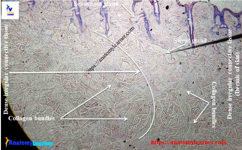
In the thin aponeuroses or fascia of muscles, the collagen bundles are located in a single layer. You will find collagen fibers in several layers in heavier aponeuroses, organs capsule, or the dermis. Generally, you will find the dense irregular connective tissue in the following organs or structures of an animal’s body.
#1. In the dermis of the thick and thin skin of animals
#2. In the fascia, aponeuroses, and joint capsule
#3. Fibrous capsules of spleen, testes, ovary, and kidney of animals
#4. Lamina propria of the digestive tract of animals.
Loose connective tissue histology
The loose connective tissue is characterized by a loose, irregular arrangement of connective tissue fibers and numerous ground substances. You might know the histology of loose connective tissue for further study of different organs or structures from the animals. I have published a simple guide to learn loose connective tissue histology with real slide pictures in anatomy learner.
Difference between two types of dense connective tissue
The main difference is the arrangement of connective tissue fibers; in irregular tissue, you will find the randomly oriented fibers bundles. Whereas fibers are oriented inconsistent patterns in dense regular tissue structure.
The cells are mainly fibroblasts in dense regular tissue, whereas you will find fibrocytes and other connective tissue cells in the dense irregular tissue. But both dense tissues have a small amount of ground substances.
Elastic connective tissue
This elastic connective tissue is the specialized dense connective tissue made of mainly elastic fibers. It may found in the place where the elasticity requires like in ligamentum nuchae.
Fibers of connective tissue
The collagen fibers occur singly or in a bundle that runs in the wavy course. Collagen fibers don’t branch and white in fresh condition. Elastic fibers appear in single and never in bundles; they branch and anastomose to form the network or framework. Reticular fibers are very thin collagen fibers and have similar histological features to collagen fibers. They form the supportive frameworks of different lymphoid organs and glands.
Drawing of dense irregular tissue slide image
I will share the drawing of dense irregular connective tissue slide images so that you might follow the same drawing.
If you need more slide images or a labeled diagram of dense connective tissue, you may follow anatomy leaner on social media (get more related or updated images of dense connective tissue slides).
You might read other articles (histology and anatomy of different organs of animals) from anatomy leaner –
#1. Histological features of the spleen with slide images and labeled diagram
#2. Thyroid gland histology slide and description
#3. Histology of pancreas with real slide pictures and labeled diagram.
Conclusion
You have learned the basic histology of dense irregular connective tissue with slide pictures and labeled diagrams. Are these identification points helpful to identify dense irregular connective tissue slides under the microscope?
If you want, you may share this article with your friends who want to know the basics of dense connective tissue histology. Stay connected, and don’t forget to read other articles from anatomy learner.