
Uterus is the site of implantation of conceptus and undergoes a definite sequence of change during estrous and reproductive cycle. In uterus histology you will find three defined layers – mucosa submucosa (endometrium), muscularis (myometrium) and serosa (perimetrium).
Hi dear, do you looking for the best guide to learn uterus histology with slide pictures and labeled diagram? Fine, in this article I am going to describe the histological features of different layers of uterus. I will show you the every single structure from uterus histology slide with labeled pictures.
You will also find a uterus histology drawing slide image at the end of this article. If you really interest to know the histology of endometrium, myometrium and perimetrium from uterus then continue this article till end.
I hope you know the anatomy of uterus; if you want to memorize the anatomical structure of uterus then read this article from anatomy learner.
Okay, let’s get into the main part of the article – uterus histology with real slide picture and labeled diagram.
Uterus histology
First I would like to enlist the histological structure that you might identify under the light microscope from uterus histology slide at laboratory. This will help you to learn the details histological features of uterus later.
From endometrium of uterus histology you might identify the following structures –
#1. Lining epithelium (simple columnar epithelium)
#2. Thick lamina propria of endometrium
#3. Uterine gland with their lining epithelium (branched and coiled; varies on condition)
#4. Structure of functional layer and basal layer of endometrium (you will learn details on endometrium part)
You might identify the flowing structures from the myometrium layer of uterus –
#1. Longitudinal smooth muscles layer
#2. Cross section of smooth muscle layer in myometrium of uterus
#3. Intestinal connective tissue of myometrium layer
#4. Blood vessels on myometrium layer
You might also identify the simple squamous lining epithelium from outer serous coat of perimetrium.
Identification points of uterus histology slide
These are the most important identification points of uterus histology slide under light microscope. Let’s try to identify uterus slide under light microscope with these identifying characteristics –
#1. Presence of inner endometrium, middle myometrium and outer perimetrium layers
#2. Endometrium (thicker) is lined by simple columnar epithelium and contains more cells and uterine glands (branched and coiled glands in secretory phage)
#3. Presence of thick myometrium that having a thick middle circular layer of smooth muscle bundle with numerous blood vessels
#4. The outer perimetirum is lined by simple squamous epithelium or mesothelium
So this is the uterus slide of animal. Okay, let’s find these characteristics from the uterus slide picture.
Uterus histology slide with labeled diagram
Do you want to know the details histology of different layers of uterus with real slide and labeled diagram? Here I am going to describe the histological characteristics of three different layers of uterus from animals.
#1. Endometrium layer histology
#2. Myometrium layer of uterus
#3. Perimetrium layer histological features
Endometrium histology with slide picture
In endometrium histology you will find two zones – superficial functional zone and thin deep layer called basal zone. The superficial layer of endometrium layer degenerate partially or completely during a reproductive, estrous or menstrual cycle.
You will find the simple columnar epithelium lining on the surface of superficial zone of endometrium histology of uterus in most of the animals like mare, dog and queen. But in sow and ruminant you will find pseudostratified columnar epitheliumor cuboidal epithelium lining.
Again the functional zone of endometrium contains loose connective tissue with higher number of cells (mainly fibroblasts, macrophages and mast cells). The deep part of endometrium consists of loose connective tissue with less numbers of cells.
There are simple coiled and branched tubular uterine gland are found in the functional and basal zone of endometrium layers of uterus. These uterine glands are lined by simple columnar epithelium. The growth and branching enhanced by rising estrogen level.
The branching and coiling of uterine glands are more in mare and less in carnivorous animals. Again in proliferative phage of uterus you will find less, simple straight tubular gland with narrow lumen. But in secretory phage of uterus you will find more, highly coiled uterine gland with dilated lumen.
Myometrium of uterus
Myometrium is the thickest middle layer of uterus histology. This layer is consists of mostly inner circular and outer longitudinal layer of smooth muscles. In pregnancy, the number and size of this myometrium layer may increase. You will also find large arteries, vein and lymphatic vessels in this myometrium layer of uterus.
Due to presence of larger numbers of vessels in the myometrium layer, it is also known as stratum vasculare.
Perimetrium layer of uterus
In uterus histology, you will find the outer serous coat which is known as the perimetrium. This perimetrium layer of uterus has simple squamous epithelium lining (known as mesothelium) that covers the loose connective tissue. You may also find the smooth muscles cells in the perimetrium layer of uterus. This layer is continuous with the structure of the broad ligament of uterus.
Proliferative and secretory phage of uterus
How you will differentiate proliferative and secretory phage of uterus histology slide? Well, I will enlist few points that will help you to different the proliferative uterus structure from secretory uterus of animals.
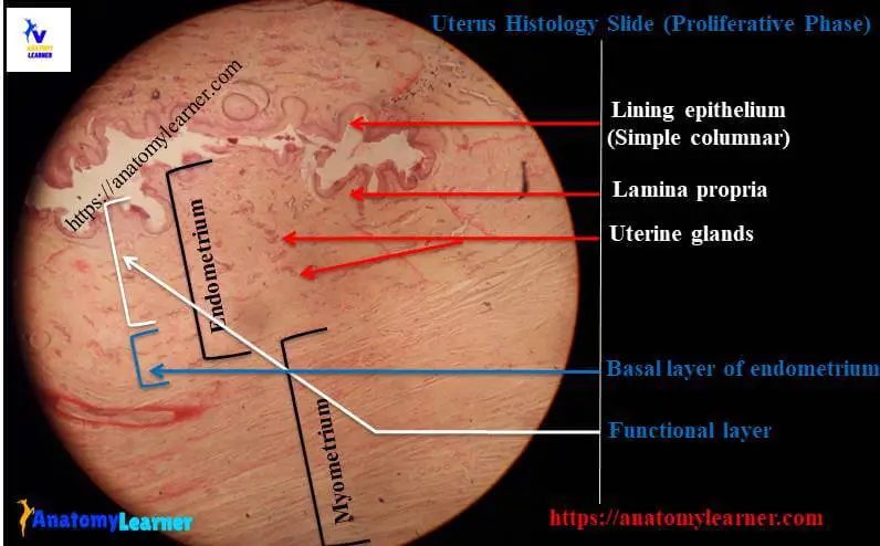
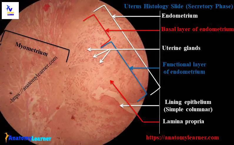
#1. The thickness of endometrium layer is more in secretory phage of uterus compare to proliferative phage
#2. You will also find difference in the lining epithelium in two phages of uterus; the lining epithelium of proliferative phage is simple columnar and hypertrophied columnar epithelium in secretory phage
#3. There are less uterine glands (simple straight tubular) with narrow lumen in proliferative phage; but in secretory phage you will find the more, highly coiled uterine gland with dilated lumen.
#4. No secretory material will find in the lumen of proliferative phage; but in secretory phage – there may present secretion in the lumen of glands.
#5. The stroma is highly cellular and non-edematous in proliferative phage, whereas stroma is highly vascular and edematous in secretory phage.
Hope these points will help you to identify the proliferative and secretory phage of uterus.
Uterus histology drawing pictures
Now I am going to share the simple uterus histology drawing pictures with you. You might try to draw the better uterus pictures.
If you need more pictures related to uterus slide and labeled diagram then you may follow anatomy learner at social medial for more update on pictures.
You might like other article related to female genital organs – ovary histology with labeled diagram, fallopian tube or uterine tube histology from anatomy learner.
Conclusion
I hope you got the best guide to learn uterus histology with real slide picture and labeled diagram from anatomy learner. Hope the main identifying characteristics of uterus slide histology will help you to identify the proliferative and secretory phages of uterus.
If you think this article is helpful and informative for learning the histological features of uterus then you may share this article with your friends who want to learn layers of uterus.