
Do you want to learn the ureter histology with anatomy learner? In ureter histology you will find three major layers under the light microscope.
You know the ureter is a muscular tube like structure and convey urine from the renal pelvis of each kidney to the urinary bladder. The peristaltic movement of smooth muscles layers of ureter wall helps to conduct urine from pelvis to urinary bladder.
In this article, I am going to share the basic ureter histology layers with real slide pictures. I will also show you the ureter histology labeled diagram and drawing.
Here you are going to get a full ureter histology guide where you will learn all histological features of a ureter.
You may also get little information about urethra histology, urinary bladder histology and kidney histology at the end of this article. If you want you may learn details histology of the above organs from anatomy learner blog.
Okay, let’s get into the main part of the article – ureter histology with slide pictures and labeled diagram.
Ureter histology
Hope you know the basic organizational pattern of a tubular organ. If you want to memorize the histological features of a tubular organ then you may read this article. Hope this organizational patter of the tubular organ will help you to understand the histological features of ureter easily.
In ureter histology you might find the following important histological characteristics under the light microscope –
#1. The epithelium (transitional epithelium) lining of tunica mucosa of ureter in animal
#2. Two or three layers of smooth muscles at tunica muscularis in different animals
#3. Fibro-elastic adventitia layer at outer part of ureter in animal
#4. Star or stellate shaped lumen of ureter of animal
Fine, now you might find these structures from the labeled real histological pictures below. Hope you will understand the histological features of ureter of animals.
Ureter histology layers
So, what are the main layers found in ureter? Okay, you will find the following three main ureter histology layers –
#1. An inner epithelium lining of mucosa membrane or tunica mucosa
#2. Middle smooth muscles layers (two or three muscle layers) or tunica muscularis
#3. Outer fibro-elastic adventitia or tunica adventitia layer
Want to learn more about these ureter histology layers? Fine, let’s continue this article to know the different layers of ureter from different animals.
Tunica adventitia of animal ureter
The ureter of different animals have narrow lumen; if you don’t know the ureter anatomy then please read this article from anatomy learner blog.
In normal condition, the longitudinal folds of tunica mucosa provide the lumen a stellate shape appearance in cross section.
Do you know how these longitudinal folds are formed in ureter wall? These folds created from the looseness of the outer layer of lamina propria. You will find this stellate shaped ureter lumen in normal condition. But in distended condition of ureter, these folds of mucosa will disappear.
The mucosa membrane of ureter consists of transitional epithelium lining with five to eight cell layers. This transitional epithelium is supported by underlying loose connective tissue layer called lamina propria. The lamina propria of ureter histology is rich in elastic tissue. There is no distinct sub-mucosa layer in ureter histology.
In horse ureter, you may find the mucosal gland at the proximal part of ureter.
Tunica muscularis layer of animal ureter
There is thick tunica muscularis layer in ureter structure which consists of bundles of smooth muscle cells. These smooth muscle bundles are arranged in two different patterns in most of the ureters of different animals –
#1. Inner longitudinal smooth muscle bundles layer in ureter and
#2. Outer circular layer of smooth muscle bundles in ureter
But sometime these two layers of smooth muscle bundles are difficult to distinguish in ureter of few animals.
At the lower one third of ureter histology of animal, you might find another smooth muscle layer. This third smooth muscle bundles layer oriented longitudinally and present at outside of the circular smooth muscle bundles.
So, at lower one third of ureter you will find three smooth muscle bundles at tunica muscularis layer –
#1. Inner longitudinal smooth muscle bundle in ureter
#2. Middle circular smooth muscle bundle layer in ureter and
#3. Outer longitudinal smooth muscle bundle of ureter
Tunica adventitia of animal ureter
Tunica adventitia is the outer layer just external to tunica muscularis layer of ureter histology. In ureter you will find fibro-elastic connective tissue in tunica adventitia.
You will also find numerous blood vessels and fat cells in tunica adventitia of ureter.
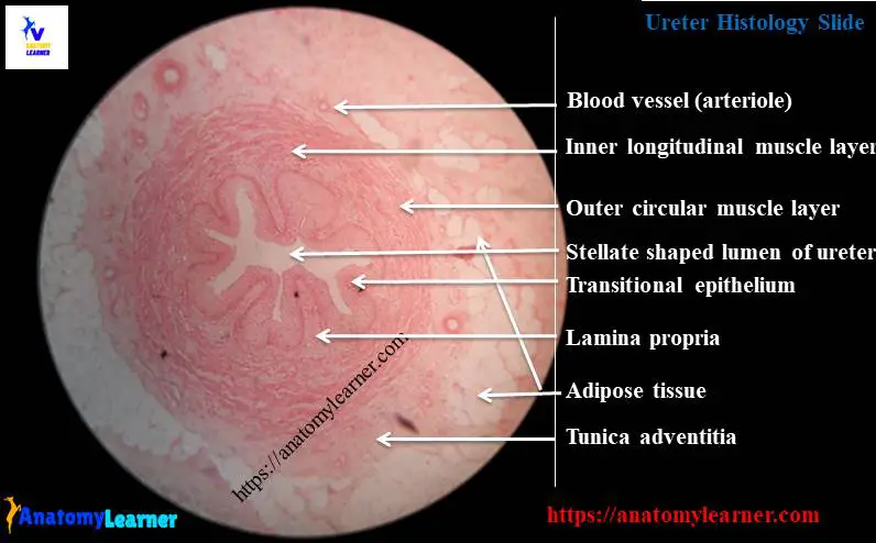
Ureter histology labeled diagram
From this ureter histology labeled diagram you will able to identify all the important histological structures. If you want to more ureter slide pictures and labeled diagram I would like to invite you to join anatomy learner at social media. You will get updated ureter slide pictures and labeled diagram.
How to identify ureter slide under light microscope?
Hope, following important ureter slide identifying histological characteristics will help you to identify the ureter structure under light microscope
#1. Presence of mucosal folds which is lined by transitional epithelium
#2. Stellate shaped lumen (of ureter might found in normal condition)
#3. Presence of inner longitudinal smooth muscle layer and outer circular smooth muscle layer in ureter
#4. More fat cells and blood vessels are found in tunica adventitia of ureter
Urethra vs ureter histology
Do you want to learn the difference between urethra and ureter histology? You will get a details guide about urethra histology article at here in anatomy learner blog. After reading that guide you will able to make the difference between urethra and ureter histology.
Urinary bladder histology
You will find the same histological features in urinary bladder histology like ureter or urethra. But there are some special histological features that are requires to known to identify the urinary bladder under light microscope. If you are interest to learn urinary bladder histology then you might find the article at here in anatomy learner blog.
Conclusion
Hope you got a better guide to learn ureter histology with labeled diagram from anatomy learner. Now you could able to identify the ureter slide under light microscope with proper identifying characteristics.
If you found any mistake at ureters histology labeled diagram then please let me inform. Again, I will request you to learn the general organizational pattern of a tubular organ properly so that you might understand the ureter histology layers.
If you found any value then I would like to request you to share this article with your friends who want to get better ureter histology guide. Read other article like urinary bladder histology and kidney histology from anatomy learner.