
In fallopian tube histology, you will find little variation in the different parts of fallopian tube of animal. It is so easy to describe the basic histology of fallopian tube of animal if you have the knowledge of general organizational pattern of any tubular organ.
Hello dear, do you want to learn the fallopian tube histology with real slide pictures and labeled diagram? Fine; in this article I am going to share the basic histological structure of a fallopian tube from animals.
You will learn the little difference in different parts of fallopian tube histology from this article. I hope, after completing this article you will able to identify the different parts of fallopian tube slide under light microscope.
You might know the fallopian tube anatomy first to start learning histological features of fallopian tube. If you want you might read this article where you will find the fallopian tube anatomy at the middle part of article.
Again, I will highly recommend you to learn hollow organ histology before to start learning fallopian tube or uterine tube histology of animals.
Fallopian tube histology
I would like to enlist the most important identifying histological features from fallopian tube histology slide under light microscope. Then I will go details histology of each parts of fallopian tube in animal.
#1. Presence of highly folded mucosa (varies in different parts) which is lined by the simple columnar or pseudostratified columnar with motile cilia
#2. The lamina propria of fallopian tube contain more numbers of cells
#3. Presence of thick inner circular layer of smooth muscles and thin outer longitudinal layer of smooth muscle in tunica muscularis
#4. Serosa of fallopian tube consists of mesothelium supported by connective tissue
Hope, you got the most important identifying histological characteristics of fallopian tube under light microscope. Now you might learn the details histology of uterine tube from animal.
Fallopian tube anatomy
I am going to the overview of fallopian tube anatomy from animals. You know fallopian tubes are bilateral and tortuous structure that extends from ovary to uterine horn. You will find mainly the three distinguished fallopian tube parts in animal –
#1. Infundibulum of fallopian tube in animal
#2. Ampulla part of fallopian tube and
#3. Isthmus part of fallopian tube
Infundibulum is the larger funnel shaped structure in fallopian tube anatomy. Ampulla is the thin walled structure just behind the infundibulum part of fallopian tube. Again, isthmus is the narrow muscular joining segment between fallopian tube and uterus.
Okay, if you want to know more about the anatomy of fallopian tube or uterine tube or oviduct of animal, then you might learn from the veterinary gross anatomy section.
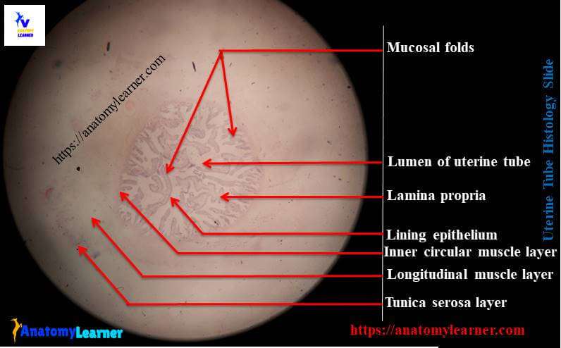
Fallopian tube histology with slide and labeled diagram
Okay, let’s know the details histology of fallopian tube with real slide and labeled diagram. In fallopian histology you will find the following layers or structures –
#1. Mucosa of fallopian tube with lining epithelium
#2. Propria submucosa of fallopian tube in animal
#3. Tunica muscularis layer of fallopian tube and
#4. Tunica serosa layer of fallopian tube
Fallopian tube epithelium histology
You know the mucosa of fallopian tube have many longitudinal folds; but the numbers of folds may varies with animals and also in different parts of fallopian tube. The mucosal folds are more in ampulla and gradually decrease at the isthmus part of fallopian tube.
You will find more longitudinal folds of mucosa in the ampulla of mares and sow. There are moderate numbers of mucosal folds found in fallopian tube of cow; again these folds branched into secondary and tertiary folds.
Okay, now identify the fallopian tube epithelium histology. You will find the simple columnar or pseudostratified columnar epithelium with or without cilia at the mucosal lining of fallopian tube. In the cranial part of fallopian tube you will find more numbers of ciliated and secretory cells.
Propria submucosa of uterine tube
In most of the organs from female reproductive system of animal lacks of lamina muscularis layer. So, the lamina propria continuous with submucosa in fallopian tube
You will find loose connective tissue along with different types of cells like mast cells, plasma cells in the propria submucosa of fallopian tube.
The submucosa of ampulla is more folded than the other parts of fallopian tube in animals; gradually this folds decrease to isthmus part.
Tunica muscularis and serosa layers of fallopian tube
In most of the animals, you will find a thick circular smooth muscle layer in the tunica muscularis layer of fallopian tube. But you may also find other different arrangement pattern of smooth muscle layers like – isolated longitudinal and oblique bundles.
In infundibulum of fallopian tube, you will find the two layers of smooth muscles bundle – inner circular and outer longitudinal layer. These muscle layers are thin in infundibulum part and thick in ampulla of fallopian tube. In isthmus part of fallopian tube the inner circular smooth muscle layer is thicker than other parts.
The serosa of fallopian tube is consist of loose connective tissue and lined by endothelium.
Fallopian tube histology drawing
I am going to share my fallopian tube histology drawing with you. Hope this fallopian tube slide drawing will help you a lot.
Please try to draw better fallopian tube histological structures than this drawing.
If you need more drawing of fallopian tube histology then you may follow anatomy learner at here in social media. You will also get more real slide picture and different labeled diagram of different organs from animal body.
You might also learn the histological features of different organs or structures from female genital system of animal –
#1. Histological features of ovary, corpus luteum of animal
#2. Uterus histology – different layers of animal uterus
#3. Histological characteristics of mammary gland of animal
Again, if you want to learn histology from other different organs systems then you may follow anatomy learner histology learning section.
Conclusion
I hope you learn the basic fallopian tube histology with anatomy learner. Now you will able to identify the fallopian histology slide under light microscope.
Do you like this fallopian tube histology labeled diagram? Fine, if you think this article was helpful then you may share this article with your friends who want to learn histological features of different organs from female genital system.
Don’t forget to read the latest article from anatomy learner from here.