
In pancreas histology of animal, you will find the both endocrine and exocrine parts. The exocrine part forms the major portion of pancreas and consists of closely packed serous acini with zymogenic cells. Again, the endocrine part consists of pancreatic islets of Langerhans which is located within the masses of serous acini.
Hi, do you want to learn pancreases histology with real slide images and labeled diagram? In this article I am going to share the basic histology of pancreas along with their identifying characteristics under light microscope.
First I will show you the different important structures or components from pancreas histology slide like – pancreatic acini, islets of Langerhans then I will go with the details description of pancreas histology.
If you really interest to learn the histological features of exocrine and endocrine part of pancreas then you might continue this article.
Pancreas histology
You know pancreas is an encapsulated, lobulated and compound tubuloacinar gland in animal. From the pancreas histology, you might identify the following histological structures –
#1. Capsule of pancreas
#2. Interlobular connective tissue septa of pancreas
#3. Serous acini with zymogenic cells of exocrine part
#4. Interlobular ducts of pancreas
#5. Intercalated ducts of pancreas structure
#6. Centroacinar cells of serous acini of pancreas
#7. Pancreatic islet of Langerhans of pancreas (endocrine part)
#8. Blood vessel on the interlobular connective tissue septa
#9. Pacinian corpuscle of pancreas structure (optional)
Okay, let’s find these important histological features from the pancreas histology real slide and labeled diagram. Hope this pancreas histology images will help you to understand the components of pancreas.
Identifying features of pancreas histology real slide
You might identify the pancreas histology real slide under the light microscope in laboratory. Hope, the following important identifying characteristics will help you a lot to identify pancreas structure under microscope.
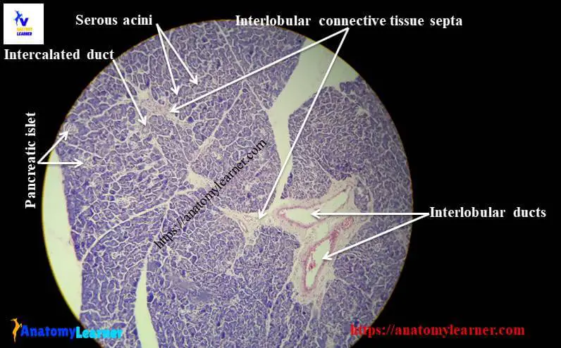
#1. Presence of darkly stained closely packed serous acini arranged in small lobules of pancreas
#2. The small lobules are surrounded by the intralobular and interlobular connective tissue septa that contain blood vessels and interlobular ducts.
#3. Presence centroacinar cells in the middle of the serous acini of pancreas
#4. Highly stained pancreatic islets of Langerhans are present within the masses of serous acini of pancreas histology structure (most characteristic feature of pancrease).
Hope you will identify pancreas histology slide under light microscope with these important identifying characteristics.
Pancreas histology description
So, do you want to learn the details description of pancreas histology? If you want then you might continue this article to learn more about pancreas structures.
You know there is two main parts of pancreas – exocrine part (serous acini) and endocrine part (islet of Langerhans). I am going to describe the histological features of these two parts from pancreas structure separately.
Pancreatic acini histology
You know, in any glandular structure you will find these two main structures – stroma and parenchyma. The stroma of pancreas consists of a thin capsule and this capsule gives rise a delicate connective tissue septa that separate the parenchyma into different distinct lobules.
The exocrine part of pancreas form the major part of pancreas and you will find the densely packed serous acini that arranged into many lobules.
The lobules of exocrine pancreas are separated by the interlobular and intralobular connective tissue septa. You will find the blood vessels, interlobular ducts and occasionally sensory receptor pacinian corpuscle on these connective tissue septa of pancreas histology structure.
In pancreatic acini, you will find pyramidal shaped cells (contains spherical nucleus; located at the base of cell) that surrounding a small lumen. The cytoplasm of pyramidal cells of pancreatic acini is intensely basophilic and contains more number of rough endoplasamic reticulum and mitochondria. The apical part of these pancreatic acini cells contains the eosinophilic membrane bounded zymogen granule.
The small lumen of pancreatic acini cells continuous with an intercalated duct. This duct begins with flattened cells that invagineted into the acinar lumen; this cells is known as the centroacinar cells.
The intercalated ducts of pancreas structure joins with intralobular ducts which are lined by simple cuboidal epithelium. Again, this intralobular duct continues with interlobular and collecting ducts that are lined by the columnar epithelium.
Pancreatic Islets of Langerhans histology
In pancreas histology you might find the most characteristics features – the pancreatic islets of Langerhans. The pancreatic islets of Langerhans are the pale staining ovoid masses or bodies (structure may vary; generally ovoid or spherical shaped) among the serous acini of pancreas structure.
The endocrine cells are larger than serous acini cells and cluster in pancreatic islets. You may find the islets cell arranged in an irregular cord and composed of five different cells – alpha cells, beta cells, delta cells, F cells.
Alpha cells are the larger cells with eosinophilic granule of islets of Langerhans; you will find these alpha cells in the center of islets in horse, but at periphery in cow. Sometime the alpha cells lack in dog’s pancreatic islets. These alpha cells are responsible to synthesize glucagon that increases the glucose in blood level.
Beta cells of islets are small in size and have basophilic granules. These beta cells are mostly finding in the center of each pancreatic islets of pancreas. They are responsible for synthesize and store of insulin that decrease glucose level in blood.
Delta cells are few in number in pancreatic islets of pancreas histology. They are responsible for secreting somatostatin that inhibits glucogon, insulin and growth hormone. Again the F cells secrets pancreatic polypeptide that inhibits pancreatic secretion.
Pancreas histology slide labeled diagram
Here I am going to share few pancreas histology real slide and labeled diagram with you so that you might understand and learn the histological structures of pancreas completely. If you need more images related to pancreas real slide then you may follow anatomy learner at here in social media. You will also get updated pictures and article notification at here.
If you found any mistake in labeled diagram of pancreas images then let me inform. I will highly appreciate your support to enrich histology learning.
Pancreas anatomy
If you want to learn pancreas anatomy then you may learn with anatomy learner at here (veterinary gross anatomy section).
Conclusion
This is the short guide to learn the histological features of pancreas. Hope you will able to identify pancreas histology real slide under light microscope. Do you like these pancreas histology slide images and labeled diagram? If you found they useful then share this article with your friends who want to know the histological features of pancreas with short but perfect guide.
Don’t forget to read the other different article related to histology of different organs from anatomy learner histology learning section.