
Thymus of animal is a central lymphoid organ which is essential for the growth and development of other lymphoid organs. In thymus histology you will find the dark staining outer cortex and light staining inner medulla.
Hi, do you want to identify normal thymus histology slide under the light microscope with proper identifying characteristics? In this article I am going to share the basic thymus histology description with real slide and labeled diagram with you. You will also get the possible important identifying characteristics of thymus gland structure so that you might identify thymus slide easily.
I will share my hand drawing thymus histology slide picture with you; hope this will help you a lot to understand all the histological structures of thymus from animal. After completing this article you will able to identify the histological features from thymus slide.
So, if you are really interest to learn the normal thymus gland histology with best information, labeled slide images and video then you may continue this article.
If you want to know the other histological features of different organs from animal body then you may read all the latest articles from anatomy learner histology learning section.
Okay, let’s get into today’s article – normal thymus gland histology with slide labeled diagram
Thymus histology
Thymus of animal is a bilobed organ where each lobe being equal in size. It has dual origin and known as the lympho-epithelial organ of the body. The lymphocytes of thymus structure originated from mesoderm whereas the reticular epithelium cells originated from endoderm of pharyngeal pouch.
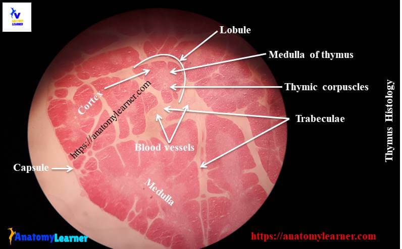
From the thymus histology slide you might identify the following important structures under the light microscope at laboratory.
#1. Thin connective tissue capsule of thymus structure
#2. Interlobular connective tissue septa or trabeculae of thymus
#3. Dark staining outer cortex of thymus
#4. Light staining inner medulla of thymus structure
#5. Numerous incomplete lobules of thymus
#6. Thymic or Hassall’s corpuscles at the medulla of thymus structure
#7. Blood vessels at capsule and thin trabecuale of connective tissue
#8. Adipose tissue both in thymic lobules and connective tissue trabeculae
Now, try to find out these histological features of thymus from the labeled diagram. Hope these will help you a lot to understand thymus structure.
Thymus histology slide identification
I am going to enlist the most relevant important histological features that might help you to identify the thymus histology slide under light microscope.
#1. Presence of many incomplete lobules that are partially separated from the other lobules by thin connective tissue septa or trabecuale
#2. There are darkly stained cortex and lightly stained medulla present in each lobule of thymus structure
#3. Presence of densely packed medium sized lymphocytes in the cortex of thymus
#4. Hassall’s corpuscles are present in the medulla of thymus structure
#5. Presence of numerous blood vessels and adipose cells in both the thymic lobules and the connective tissue septa or trabeculae
Hope, you could identify thymus slide under light microscope with these identifying characteristics.
Thymus histology description with labeled diagram
In thymus histology you might describe the supporting framework (capsule, stroma) and the lobules or parenchyma.
A thin connective tissue capsule enclosed the thymus structure completely. This connective tissue capsule continuous with the septa that subdivides the lobe into partially separated lobules.
In each lobule of thymus you will find the outer cortex and inner medulla. I am going to describe the cortex and medulla of thymus separately.
Cortex of thymus structure
The thymic cortex is consists of mainly the lymphocytes (T lymphocytes) and the epithelium reticulum. The reticulum is stellate shaped and have large ovoid, pale nuclei and long branching cytoplasmic process. These cytoplasmic processes of epithelium reticulum come in contact with the other reticulum epithelial. The epithelial reticulum forms the supporting stroma of the thymus histology.
The cytoplasm of these epithelial reticulum contains the secretory granules that help to secrete different hormone. You will find lymphoblast and medium size lymphocytes in the outer part of the cortex of thymus structure. These medium sized lymphocytes divide by mitosis to produce the smaller lymphocytes which are pushed into the deeper part of cortex.
You will find few macrophages in the cortex of thymus structure. The cortex of thymus is darker because it contains number lymphocytes.
Medulla of thymus structure
The medulla of thymus histology contains fewer lymphocytes and more epithelial reticular cells. Here you will find the most characteristic features and that is Hassall’s corpuscles or thymic corpuscles.
The Hassall’s corpuscles are the ovoid or round lamellate acidophilic bodies that have a central homogenous hyaline material surrounding by concentric layers of flatten epithelial cells.
Thymus is generally active in young animal and reaches its peck development at puberty. After that the involution may occurs in thymus structure. Gradual depletion of lymphocytes is the most characterize feature of thymus involution.
This depletion of lymphocytes mainly occurs in the cortex of thymus. The epithelial reticular cell become enlarges and the parenchyma is invaded by white adipose cells.
Thymus histology drawing
I am so excited to share thymus histology drawing pictures with you so that you might understand the every single part of thymus easily. You might try to draw the better thymus slide structure. If you need more update thymus slide pictures then you may follow anatomy learner at here in social media.
If you find any mistake in thymus histology drawing then please let me inform. I will highly appreciate your kind support to make thymus learning easy for all.
Learn histological features of other organs
You might also learn the histological features of different organs from lymphoid tissue –
#1. Histological feature of lymph node with real slide pictures
#2. Identifying histological features of different tonsil of animal
#3. Histology of spleen – the white and red pulp structure
Conclusion
Hope you got the best guide to learn the basic of thymus histology with slide and labeled diagram from anatomy learner. Now you will able to identify the thymus histology slide under light microscope.
If you think this article is helpful then you may share it with your friends who want to learn the histological features of thymus structure.
Again, don’t forget to follow anatomy learner histology learning section to get more updates articles and labeled pictures