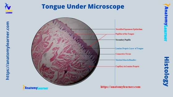
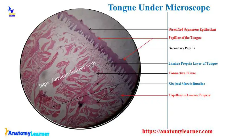
The tongue under a microscope shows a core of crisscrossing skeletal muscle bundles and a peripheral mucous membrane. A stratified squamous epithelium covers the mucous membrane of the tongue and contains 4 types of papillae.
Again, each tongue papilla possesses a connective tissue core that covers the epithelium. Some of these papillae of the animal’s tongue contain taste buds.
Finally, you will find numerous blood vessels and serous and mucous glands in the connective tissue around the skeletal muscle fibers of the tongue.
This article will help you to understand (identify) the basic structure of the tongue under the microscope with the labeled diagram. You will also get basic information of tongue histology from different species like cats, dogs, lions, and tigers.
So, if you want to learn and identify the tongue microscope slide, let’s continue this article till the end.
Tongue under microscope
The tongue is a muscular organ that covers with mucosa. And it is important in the prehension, mastication, and deglutition of feed. A tongue under a microscope shows the below-mentioned histological features –
- Nonkeratinized stratified squamous epithelium on the tongue mucous membrane,
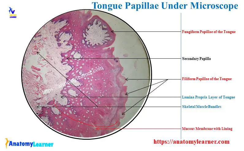
- Different types of papillae – filiform, fungiform, circumvallate, and foliate,
- Lamina propria layer in the mucosa membrane of the tongue,
- Different types of lingual glands (serous, mucous, and mixed) and their ducts in the tongue,
- The core of the crisscrossing bundle of skeletal muscle in the tongue, and
- Connective tissue, blood vessels, and nerves in the core of the tongue,
Again, you will find 3 types of cells in the taste bud of the tongue papillae (not shown in the microscope slide). Let’s see all of the above-mentioned histological features from the tongue microscope slide labeled diagram.
You will find some special lingual structures in the tongue of a dog and ruminant (cow, sheep, and goat). I will describe the special lingual features of these animal species at the end of this article.
Tongue microscope slide identification
Here, I will provide the identifying points of the tongue (basic structure), papillae (filiform, fungiform, and circumvallate), and taste buds from a microscope slide. First, let’s identify the basic lingual structure under the light microscope with the help of the followings points –
- The sample tissue section under the light microscope shows the nonkeratinized squamous epithelium on the mucous membrane,
- There are numerous mucosal projections (known as the papillae) in the sample tissue section,
- The mucous membrane shows the conical, filiform papillae (no taste bud) and mushroom-shaped fungiform papillae,
- The core of the tissue section shows skeletal muscle bundles that run in different directions,
- Numerous blood vessels, serous, and mucous glands are present amongst the skeletal muscle bundles of the sectioned tissue,
So, this is the tongue microscope slide (most of the structures are identified from the actual tongue microscope slide).
Tongue papillae identification
The dorsum of the tongue bears different types of microscopic lingual papillae (varies in different animal species). You may easily identify the different papillae from the tongue of various animals with the help of the below-mentioned identifying points –
- Presence of slender, threadlike (conical) shaped filiform papillae on the dorsal surface of the tongue,
- Conical papillae are larger than the filiform and present in the root of the tongue of dogs, cats, and pigs (also found in the torus lingua of a ruminant),
- Presence of flattened, lens-shaped projections (lenticular papillae) on the torus lingual of the ruminant tongue,
- Presence of dome-shaped or mushroom-shaped scattered fungiform papillae on the dorsal surface of horse and pig’s tongue, and
- A flattened and large vallate papillae present on the dorsum of the animal’s tongue (rostral to the root),
In the vallate and circumvallate papillae of the animal’s tongue, you will find some special histological features –
- It is a sunken inverted cone-shaped papilla with a flat top that lines with the stratified squamous epithelium,
- Presence of numerous taste buds on the lateral wall of the circumvallate papillae,
- There are deep, circular furrows around the circumvallate papillae of the tongue, and
- The skeletal muscle runs in different directions of the structure,
How to identify taste buds in a tongue?
You may easily identify the taste buds from the tongue with the help of the followings identification points –
- Presence of a cluster of specialized epithelial cells (spindle) embedded in the stratified squamous epithelium of the tongue’s papillae,
- They take light stains and possess spindle-shaped gustatory and sustentacular cells,
- There presence of a taste pore at the epithelial surface of the taste buds,
I hope you already got the basic idea of the general histological structure of the tongue. Now, I will summarize the microscopic features of the animal’s tongue in the next section of this article.
Tongue microscope slide description
This is a very short description of the tongue under a microscope. You know, the tongue is a muscular organ that is made of skeletal muscle. You will find the mucous membrane on the tongue’s external part.
Here, the mucous membrane of the tongue consists of keratinized stratified squamous epithelium and underlying lamina propria. The dorsum of the tongue possesses different types of mucosal projection, known as the papillae.
The number and types of papillae on the dorsal surface of the tongue vary with the animal species. In the structure of the tongue’s papillae, you will find the connective tissue core.
The histological features of the different types of papillae (like filiform, fungiform, lenticular, vallate or circumvallate, and foliate) will describe in the next part of this article.
You will find numerous slender, thread-like filiform papillae throughout the dorsal surface of the tongue. Fungiform papillae are less in number on the tongue and possess a core of the lamina propria.
In the structure of the papillae, you may find the taste bud that contains 3 different types of cells – taste or gustatory cells, sustentacular or supportive cells, and basal or stem cells.
Again, in the core of the tongue, you will find the bundles of skeletal muscles that run in different directions. So, you will find the longitudinal, transverse and oblique directions of the skeletal muscle in the section of a tongue core.
The lamina propria of the animal’s tongue possesses different types of lingual glands – serous, mucous, and seromucous (mixed). Again, the microscopic figure of the tongue shows numerous blood vessels among the skeletal muscle bundles of the core.
Now, I will describe the histology (histological features) of every single structure from the tongue with the labeled diagram.
Lingual papillae under a microscope
I hope you understand the important structures from the tongue microscope slide. If you want to describe the microscope slide of a tongue, you might point out the followings –
- The lining epithelium of the tongue (mucosa membrane) with lamina propria,
- Modification or projections of the tongue’s mucosal membrane (papillae of the tongue) with taste buds,
- Arrangement of the skeletal muscle of the tongue, and
- Lingual glands present in the tongue,
You know the lining epithelium of the tongue is the nonkeratinized stratified squamous epithelium. You may know the details of the nonkeratinized stratified squamous epithelium from the below-mentioned article of anatomy learner –
- Stratified squamous epithelium under a microscope with the labeled diagram,
Again, the lamina propria of the tongue possesses loose connective tissue and cells with some lingual glands. You may also know the details of histological features of the lamina propria from the below-mentioned article –
- Hollow organ histology with the labeled diagram (layers of a tubular organ),
Though the tongue is not a hollow organ, the microscopic features of the lamina propria are the same.
Now, the microscopic features of the skeletal muscle show the different directions in the tongue section. But, if you want to know (learn) the details and identify the skeletal muscle from any tissue, the below-mentioned article might help you a lot –
- The skeletal muscle under a microscope with the labeled diagram,
So, here, I will only focus on the microscopic features of the lingual papillae from the tongue. You will learn the microscopic features of the following tongue’s papillae with a diagram –
- Filiform papillae – slender or thread-like,
- Fungiform papillae of the tongue (dome-shaped),
- Conical papillae – conical-shaped,
- Lenticular papillae – flattened and lens-shaped), and
- Vallate and foliate papillae of the tongue,
With a diagram, let’s learn the microscopic features of these lingual papillae.
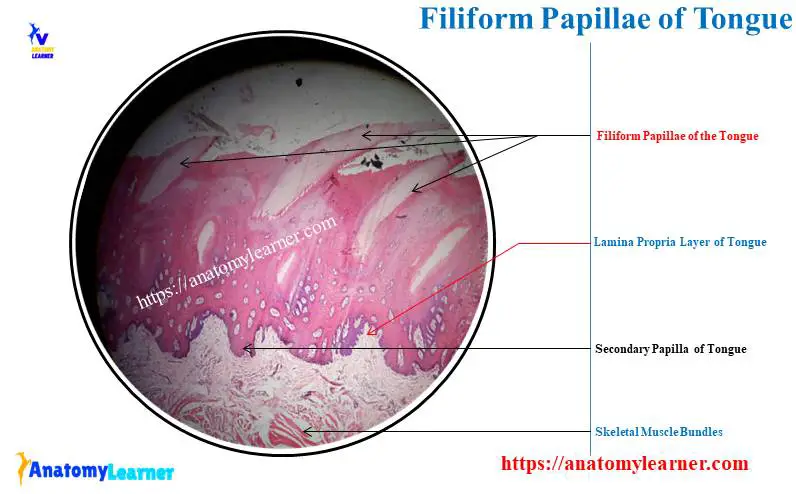
Filiform papillae of the tongue
Before describing the microscopic features of the papillae from the tongue, let’s know their type. Here, the filiform, conical, and lenticular papillae are purely mechanical. These papillae facilitate the ingesta’s movement within the animal’s oral cavity.
Again, the fungiform, vallate, and foliate papillae are gustatory and contain taste buds. These papillae of the tongue are responsible for the perception of the sense of taste.
You will find numerous slender, thread-like filiform papillae in the tongue under a light microscope. These papillae cover by keratinized stratified squamous epithelium (identified) with a thick stratum corneum.
In the core of the filiform papillae, you will find highly vascularized connective tissue under a light microscope. But, you will find a little species difference in the structure of the filiform papillae of the tongue.
In ruminants – filiform is a keratinized cone-shaped projection, and core connective tissue possesses several secondary papillae,
Horse – the filiform papillae is a very fine keratinized thread projected from the mucosal surface,
In dogs – filiform papillae have 3 or more apices, and the stratum corneum is thicker,
Cat’s filiform papillae – are larger and possess two equal prominences,
Now, let’s see the filiform papillae from the tongue microscope slide labeled diagram.
Fungiform papillae of the tongue under microscope
The fungiform papillae of the tongue are present on its dorsal surface. They remain scattered among the filiform papillae of the tongue.
Under the microscope, you will find them as the dome-shaped (mushroom) projection on the dorsum of the horse and pig tongues. A nonkeratinized stratified squamous epithelium cover surface of the fungiform papillae of the tongue.
As the fungiform papillae are the gustatory type, you will find taste buds within the lining epithelium. In the horse and cow tongue, you will find a less amount of fungiform papillae.
But, in the sheep and pig tongues, you will see more numerous fungiform papillae on their dorsum. Again, dog and goat tongues possess abundant mushroom-shaped fungiform papillae on the dorsal surface.
The core of the fungiform papillae of any animal possesses numerous connective tissue. You will find numerous blood vessels and nerves in these connective tissue cores of the fungiform papillae.
Conical and lenticular papillae of the tongue
Under the light microscope, you will see (find) the conical-shaped (larger than filiform) papillae on the root of the tongue. But where will you find these conical papillae on the animal’s tongue?
You will find the conical papillae on the root of the dogs, cats, and pigs’ tongues. Again, the torus linguae of the ruminant, you will also find the conical papillae.
In the structure of the conical papillae of the tongue, you will find both primary and secondary connective tissue prominence. You will not find any keratinization in the structure of the conical papillae of the animal’s tongue.
The structure of the conical papillae in the pigs is exceptional and possesses lymphatic tissue. They are the tonsilar papillae in the pig (collectively constitute the lingual tonsil).
On the other hand, you will see the flattened and lens-shaped projection on the torus linguae of the ruminant’s tongue. These flattened and lens-shaped projections of the tongue are the lenticular papillae.
Under the microscope, you will find the core of connective tissue and a keratinized stratified squamous epithelium in the lenticular papillae of ruminants.
Vallet and foliate papillae in a tongue
The vallate papillae are the larger and flattened structure on the dorsal surface of the tongue. They extend from the lingual surface and cover with the stratified squamous epithelium.
You will see numerous taste buds in the epithelium of the papillary side of the vallate papillae. The ducts from the serous secretory glands open into the sulcus (deep, circular furrow) at various levels.
So, the secretory products from the gland directly empty into the lingual surface. Numerous blood vessels and nerves are present in the core connective tissue of the vallate papillae.
The number of the vallate papillae significantly changes in the different animal species. You will find only 1 pair of vallate papillae in horse and pig tongue.
Dog possesses 4 – 6 pairs, whereas the cow possesses 8 – 18 pairs of vallate papillae in their tongue.
The microscopic figure of the tongue shows some parallel folds of the lingual mucosa. These are the foliate papillae on the tongue’s margin, just rostral to the palatoglossal arch.
You will also find the taste buds in the epithelium on the side of the mucosal folds of the tongue. There are no foliate papillae found in the cow tongue microscopic slide.
Though the foliate is gustatory type papillae, they are rudimentary and lack taste buds in the cat tongue.
Tongue cells under a microscope
Taste buds are ovoid structures in the tongue papillae that are made by clustering specialized epithelial cells. They are embedded (locates) in the stratified squamous epithelium of the fungiform, vallate, and foliate papillae of the animal’s tongue.
You may also find the taste buds in the soft palate, epiglottis, and other areas of the oral cavity and pharynx. The tongue under the microscope shows 3 types of cells in their taste bud –
- Type I and type II (sustentacular cells), and
- Type III or taste or gustatory cells (chemoreceptor cells),
You will also find some basal or stem cells in the structure of the taste bud of the tongue. Type I and II cells possess apical microvilli, and they project into the taste pore.
But what is the taste pore? You know the taste bud cells are spindle-shaped, extending from the basement membrane to the small opening on the epithelial surface. This opening of the taste bud is known as the taste pore.
Type I and II taste bud cells have a sustentacular or supportive role. In contrast, the taste bud type III cells are the chemoreceptors or taste cells.
Ventral surface of the tongue microscope slide
In the core of the animal’s tongue, you will find the skeletal muscle bundles that arrange in a different directions (longitudinal, transverse, and oblique). This arrangement of the skeletal muscle bundles gives the tongue extensive mobility to facilitate food movement into the animal’s oral cavity.
Now, let’s see the ventral surface of the animal’s tongue covered by the nonkeratinized stratified squamous epithelium. The structure of the mucous membrane possesses numerous capillaries and nerves.
In the structure of the skeletal muscle bundle and lamina propria, you will find seromucous lingual glands.
Cat tongue under a microscope
The basic structure of the cat tongue is almost similar to what I have already described earlier. But, you may find some little difference in the cat tongue under the microscopic view.
You will find the larger filiform papillae on the cat tongue that possess two unequal prominences. Here, the caudal prominence is large and gives rise to a caudally-directed keratinized spine.
There is a larger conical papilla on the root of the cat tongue, which is not highly keratinized. Again, the microscopic figure of the cat tongue shows numerous vallate papillae.
You will see the rudimentary foliate papillae in the structure of the cat tongue. But, these foliate papillae of the cat tongue lack taste buds.
The ventral part of the cat tongue contains adipose tissue (whereas the dog tongue contains the lyssa body).
Dog tongue under a microscope
Here, I will describe only the unique histological features of the dog tongue. Before that, you may learn the details anatomical facts of the dog tongue from another article of anatomy learners –
- Dog tongue anatomy with the labeled diagram,
The core of connective tissue, skeletal muscle bundles, and papillae possess a similar structure as I have described earlier. A most exceptional feature you will find at the ventral surface (identified) of the dog tongue is the lyssa body.
This lyssa body of the dog tongue is a cordlike structure that consists of a dense connective tissue capsule. The lyssa body of the dog tongue extends longitudinally at the midline, near the ventral surface of the apex of the dog tongue.
The microscopic structure of the dog’s lyssa body shows white adipose tissue, skeletal muscle, blood vessels, and nerves.
Ruminant and horse tongue microscopic features
On the gross anatomy of the large ruminant tongue, you will find the torus linguae on the dorsal surface. Microscopically, the mucosa of the torus linguae shows the thickened mucous membrane.
The thicker connective tissue papillae extend almost the surface of the epithelium of the ruminant tongue. A microscopic view of the ruminant tongue show scattered lenticular and conical papillae on the torus linguae of the ruminant tongue.
The exceptional microscopic feature of the horse tongue is the presence of a middorsal fibroelastic cord. In this fibroelastic cord, you will find hyaline cartilage, skeletal muscle, and white adipose tissue. This hyaline cartilage of the horse tongue structure is known as the dorsal lingual cartilage.
Tongue under microscope labeled diagram
In this article section, I will provide most of the tongue microscope slide-labeled diagrams (focusing on different important structures). First, let’s see the general structural view of the animal tongue from the first labeled diagram.
In the diagram, I tried to show the mucosal membrane with lining epithelial lining, lamina propria, inner skeletal muscle core, and lingual glands.
The diagram also shows the bundles of the skeletal muscle in different directions. Again, the tongue-labeled diagram shows the different types of papillae on the animal’s tongue.
The cat tongue labeled diagram shows the filiform papillae with a caudally directed keratinized spine arising from the caudal prominence. Again, the dog tongue labeled diagram shows the filiform papillae with caudally directed apices.
This dog tongue labeled diagram also shows the lamina propria and skeletal muscle bundles. The ruminant tongue labeled diagram shows the vallate papillae with a surrounding sulcus and taste buds.
You will find more labeled diagrams on the tongue of different species like rabbits, pigs, and lions on the social media of anatomy learners.
Conclusion
So, the tongue under a microscope shows different structures like the mucous membrane with lining epithelium, core skeletal muscle bundles, and different papillae. Again, the lamina propria of the papillae and tongue show different types of lingual glands.
The tongue from different species like cat, dog, sheep, goat, horse, and cow shows a slight variation under the microscope. All the provided labeled diagrams on the animal’s tongue might help you to get a basic idea of its microscopic features.