The ruminant stomach is very roomy and occupies nearly three- fourth of the abdominal cavity. It fills the left half of the abdominal cavity except the small space occupied by the spleen and a portion of the intestine.
Hi there, do you want to learn the ruminant stomach anatomy with a diagram? Well, you are in the right place where you will learn the anatomy of different chambers or parts of the stomach from the ruminant digestive system.
In this article, I will discuss the different chambers of the stomach of ruminant animals. After reading this guide, you will identify the different structures (external and internal) of the stomach of cows, goats, sheep, camel, and deer.
Fine, let’s get into the central part of the article – cattle, goat stomach anatomy.
Ruminant stomach anatomy (parts or chambers)
In ruminant animals, you will find four different chambers or parts in their stomach. The four compartments of the stomach are –
- #1. Rumen
- #2. Reticulum
- #3. Omasum and
- #4. Abomasum
I will try to help you identify the different parts of the ruminant stomach and their associated structures with real sample pictures. Later, I will discuss these parts of the stomach in detail. Let’s identify the following parts and structures from the goat stomach sample –
#1. Part of rumen
#2. Part of reticulum
#3. Part of omasum and
#4. Abomasum of goat stomach
#5. Longitudinal groove (both right and left) of rumen along with ruminal insula
#6. The cranial and caudal groove of the rumen
#7. The dorsal and ventral coronary groove of the rumen
#8. The dorsal sac and ventral sac of the rumen
#9. Caudodorsal and caudoventral blind sacs of rumen
#10. Atrium ventriculi (dome like vestibule)
#11. Internal infolds (shelf-like projection) of rumen
#12. Different parts of the reticulum (especially the internal honeycomb structure)
#13. Omasum and omasal laminae
#14. The neck of omasum of goat stomach
#15. Different parts of the abomasum
I hope this will provide you a basic conception of different parts of goat stomach anatomy. Now, I would like to discuss the detailed anatomy of different parts of the stomach – rumen, reticulum, omasum, and abomasum.
Rumen anatomy from ruminant stomach
The rumen is the larger muscular elongated sac that occupies most of the left half of the ruminant abdominal cavity. It extends considerably to the right of the median plane, and the long axis reaches from the point opposite the ventral part of the seventh or eighth intercostal space to the pelvic inlet.
The rumen is compressed from side to side and have the following parts –
- #1. Two surfaces of rumen – parietal and visceral surfaces
- #2. Two curvatures – dorsal and ventral curvatures and
- #3. Two extremities – cranial and caudal extremities
I hope you could identify the right and left longitudinal groove of the rumen. This will help you to understand and describe the anatomy of the rumen (my personal opinion).
The longitudinal groove (right and left) and the transverse groove (cranial and caudal) made the outer demarcation of two sacs of rumen – dorsal and ventral sacs of the rumen. Fine, come into the description of the surface anatomy of the rumen of animals.
Surfaces of the rumen (first part of the ruminant stomach)
You will find two surfaces in the rumen anatomy – parietal and visceral. The parietal surface (left) is convex and related to the diaphragm, left wall of the abdomen, and the spleen. You will find the left longitudinal groove at this surface of rumen anatomy.
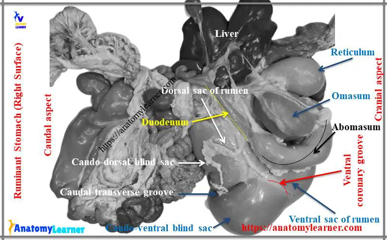
The visceral surface or right surface is irregular and related chiefly to the omasum, abomasum, intestine, liver, pancreas, left kidney, left adrenal gland, aorta, and the caudal vena cava. You will find the right longitudinal groove at this right surface of the rumen.
The right longitudinal groove extends from the cranial transverse groove to the caudal transverse groove. This right longitudinal groove encloses an elliptical area known as insula ruminis.
Again, the left longitudinal groove starts at the cranial groove, incline dorsally, then ventrally, and joins the caudal transverse groove of the rumen. It gives the dorsal branch at the midline, known as the left accessory groove.
Curvatures and extremities of goat rumen
The dorsal curvature is convex and related to the crura of the diaphragm and the sublumbar muscles. This curvature is firmly attached to these structures on the left by peritoneum and connective tissue as far caudad as the fourth lumbar vertebra. The ventral curvature of the rumen is also convex and lies on the abdominal floor.
The cranial extremity is divided into two sacs (dorsal sac and ventral sac) by a transverse cranial groove. The cranial sac is continuous caudally with the dorsal sac of the rumen, and cranially it is related to the reticulum. You will find the ruminoreticular groove externally in between the atrium ruminis and reticulum part of the ruminant stomach anatomy. The cranial extremity of the rumen is related to the diaphragm.
The caudal extremity is also divided into two sacs (caudodorsal and caudoventral blind sacs) by the deep caudal transverse groove. It extends from the pubis and is related to the intestine and urinary bladder of animals.
The caudodorsal and caudoventral blind sacs are marked off from the original sacs of rumen on each side by the dorsal and ventral coronary grooves. These coronary grooves are prominent in both the parietal and visceral surface of the ruminant rumen.
In sheep and goats, you will find a larger ventral sac than cattle and extends more to the right of median plane of animal’s body.
The internal surface of the rumen is infolded and forms a shelf-like projection known as the pillar.
Reticulum anatomy from ruminant stomach
The reticulum is the most cranial, pyriform-shaped smallest part of the four compartments of the ruminant stomach. It is located between the sixth and seventh ribs and lies on the left of the median plane of an animal’s body. Actually, you will find it in between the diaphragm and liver in the front and dorsal sac of the rumen and behind the omasum.
You will also find the following structures in the reticulum anatomy –
#1. Two surfaces of reticulum – diaphragmatic or parietal surface and visceral surface
#2. Two curvatures of reticulum (Greater and lesser curvatures) and
#3. Two extremities of the reticulum
The diaphragmatic surface is convex and lies against the diaphragm and the liver. The visceral surface of the reticulum is flattened and related to the atrium ruminis and ends dorsally by connecting the wall of the rumen. You will find the large ruminoreticular orifice in this portion of the reticulum.
The greater curvature of the reticulum faces the left and lies against the diaphragm. Again, the lesser curvature of the reticulum is located in the right dorsal and joins with the omasum.
The internal surface of the reticulum is honeycomb in appearance due to the infolding of the mucous membrane of the reticular part.
The reticulum is relatively more extensive in sheep and goats compare to an ox. The ventral part of the reticulum curves more caudally and less to the right than in cattle.
Anatomical features of the omasum
Omasum is ellipsoidal in shape and somewhat compressed laterally in ruminant. It is marked off from the other four compartments of the ruminant stomach and lies to the right of the median plane.
You will find the following structures on the anatomy of the ruminant omasum –
#1. The parietal surface of omasum
#2. The viscera; the surface of omasum
#3. Dorsal curvature of omasum
#4. Ventral curvature of omasum
#5. Base and narrow neck of omasum
#6. Reticulo-omasal and omaso-abomasal orifices
The parietal surface of the right surface of the omasum faces obliquely to the right and related to the diaphragm and liver. Ventral part of the omasum is related to the abdominal floor, right costal cartilage, and lesser curvature of abomasum.
The visceral surface or left surface of omasum faces in the opposite direction and is related to the rumen, reticulum, and abomasum parts of the ruminant stomach.
The dorsal curvatures face dorsally, caudally, and to the right. You will find a narrow neck of the omasum that is connected with reticulum in its upper part.
The reticulo-omasal and omaso-abomasal orifices are located at the upper part of the visceral surface. You will find cresent shaped flods known as laminae in the internal part of the omasum structure.
The omasum of sheep and goats is oval, smaller, and compressed laterally. It is located almost entirely to the right of the median plane.
Anatomy of abomasum
The abomasum is an elongated sac that lies on the floor of the abdomen. You will find the following parts and structures in abomasum anatomy.
#1. Funds of abomasum
#2. Body of the abomasum
#3. Pyloric part of the abomasum
#4. Visceral and parietal surfaces of abomasum
#5. Greater and lesser curvatures of abomasum
#6. Abomasal flods
Funds is the cranial blind end and located at the xiphoid region and related to rumen, reticulum, omasum, and liver. The body of abomasum extends caudally between the ventral sac of rumen and omasum. The pyloric part turns upward along the caudal border of osamus.
The parietal surface of abomasum related to the abdominal floor, and the visceral surface related to the rumen and omasum. Greater curvature provides the attachment to the superficial wall of the greater omentum, whereas the lesser curvature provides the attachment to the lesser omentum. You will also find the abomasal folds in the internal structure of the abomasum of the goat or other ruminant stomach.
Ruminant stomach diagram
I know you enjoy and learn different parts of goat stomach with real pictures. Now, I will share the ruminant stomach diagram to understand every aspect of the stomach clearly.
If you need more real pictures or diagrams of stomach anatomy then, join anatomy learner on social media.
Conclusion
I hope this easy guide will help you to learn ruminant stomach anatomy with real pictures and diagrams. If you think this is a handy guide to learn the stomach anatomy of animals, then share it with your friends who want to start learning digestive system organs anatomy from animals.
Don’t forget to join anatomy learner on social media to get more updates on articles and images.