According to the different body regions, animal vertebrae are classified into cervical, thoracic, lumbar, sacrum and caudal vertebrae. In this article, I will show you the animal vertebrae identification with labeled images.
Hi there, welcome back to anatomy learner and thanks for getting into this animal vertebrae identification article (animal bone identification). If you want to know how to identify a vertebra, then this article is for you. After finishing this article, you will identify every single part or structure from different types of vertebrae of an animal’s body.
But, make sure you know every single structure from a typical vertebra of an animal. Okay, let’s get into the main part of today’s article.
Animal vertebrae identification
This article will only focus on animal vertebrae identification. If you want to learn details on different vertebrae from the animal body, go to the specific article on anatomy learner. I will show you the different structures from the following vertebrae –
- #1. Cervical vertebrae of the animal
- #2. Identification of thoracic vertebrae of animals
- #3. Lumbar vertebrae of animals
- #4. Sacrum vertebrae of animals and
- #5. Caudal or coccygeal vertebrae of the animal
Let’s find out the special osteological features of these different vertebrae of animals.
Animal cervical vertebrae identification
The cervical vertebrae in most of the animals are seven in number. The first cervical vertebra is known as the atlas, and the second cervical is known as the axis vertebra. They are highly modified in conformity for the special function of support and movement of the head of animals.
The sixth and seventh cervical vertebrae are quadrangular and very massive. Other cervical (third, fourth and fifth) vertebrae have a long body with a median ventral crest and flat central area on the dorsal surface. Cranial extremity of these cervical vertebrae has an oval articular surface, and the caudal extremity has a nearly circular cotyloid cavity. You will find articular processes, transverse processes and spinous processes in these cervical vertebrae.
Fine, let’s identify the atlas, axis, sixth and seventh cervical vertebrae with labeled pictures.
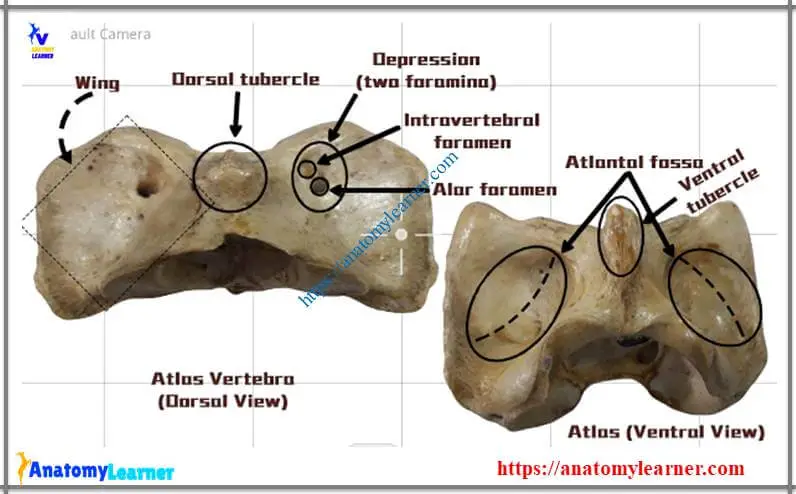
Animal atlas vertebra anatomy
The following identifying points will help you to identify animal atlas vertebra –
#1. There are no body and spinous processes in animal atlas vertebra
#2. A strong ring has formed that encloses the very large vertebral foramen
#3. There are two curved plates or transverse processes or wing that originated from the ring of the vertebra
#4. Two deep and oval cranial articular cavity are present that are separated by wide notch dorsally and a narrow notch ventrally
#5. The caudal articular surface is somewhat saddle-shaped
#6. You will find median dorsal tubercles on the dorsal arch of the atlas vertebra
#7. The ventral arch is thick, narrow and less curved
#8. There are caudal transverse concave articular surface (known as fovea dentis) on the dorsal surface of the ventral arch of the atlas
#9. You will find a modified transverse process (known as wing) that bears intervertebral foramen (medially) and alar foramen (laterally) on the dorsal surface.
#10. Again, you will find the atlantal fossa on the ventral surface of the atlas wing.
Are these identification points enough to identify animal atlas vertebra?
Identification of axis vertebra
In animal axis vertebrae identification, you might identify the osteological features of the body, arch and processes.
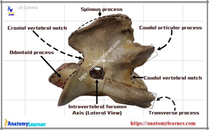
You know, the animal axis vertebra is also highly modified in conformity for special functions. Following are the important osteological features that might help you to identify animal axis –
#1. There are dens or odontoid process in the cranial extremity of the body of axis vertebra
#2. There are notch on each side of the cranial border of the arch
#3. Cranial articular process is absent whereas, caudal articular process is typical and backward directed.
#4. The transverse process is small, single (undivided) and projected caudally.
#5. The spinous process is very large and strong; the free border is rough and thickens caudally.
Sixth and seventh cervical vertebrae anatomy
In the sixth cervical vertebrae, you will find the following osteological features that will help you to differentiate it from the seventh cervical vertebrae of animals.
#1. The body of the sixth cervical vertebra is shorter and wider
#2. Arch is large and caudally
#3. The articular processes and spinous processes are well developed in the sixth cervical vertebra.
#4. Transverse processes are divided into two branches in cattle (three branches in a horse) – dorsal and ventral parts. The ventral part is a quadrilateral plate-like bone in the sixth cervical vertebra.
#5. Presence of large foramen transversarium
#6. The ventral crest is small and less prominent caudally in the sixth cervical vertebra.
But in the seventh cervical vertebra, you will find the following important osteological features –
#1. The body is shorter, wider, flattened dorsoventrally that bears facet (fovea costalis caudalis) on each side
#2. The notches on the pedicle are larger in the seventh cervical vertebra in comparison to sixth cervical.
#3. Spinous processes are well developed than the spinous processes of sixth cervical vertebra.
#4. The transverse processes of seventh cervical are not divided as you found in sixth cervical vertebra.
#5. A transverse foramen is absent in seventh cervical vertebra.
#6. The ventral crest is replaced by a pair of tubercles in seventh cervical vertebra.
I think you could differentiate the main osteological features of sixth cervical from the seventh cervical vertebrae of animals.
Animal thoracic vertebrae identification
Long spinous processes are the characteristic features in the thoracic vertebrae of animals. But, you must know the other different special osteological features for animal thoracic vertebrae identification.
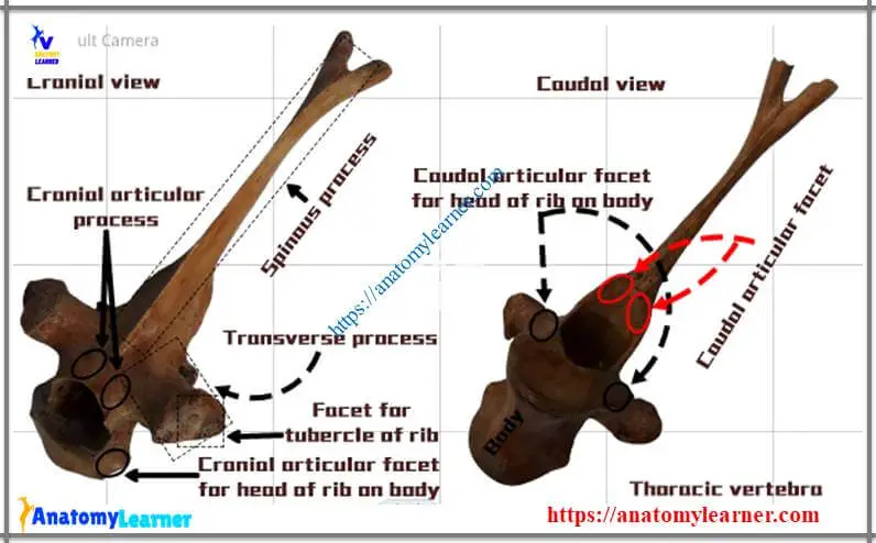
Following are the important osteological features of thoracic vertebrae that help you to identify these vertebrae from others –
#1. Presence of cylindrical, short, constricted body in thoracic vertebrae of animals.
#2. The end of the body expands and posses cranial articular facet and caudal articular facets.
#3. The arch is small, and caudal notch is relatively larger in thoracic vertebrae.
#4. Articular processes are small and underdeveloped. Cranially, you will find two oval facets that face directly dorsad. Caudal facets face almost directed ventrally in thoracic vertebrae.
#5. Transverse processes are short, thick, undivided and tuberous at the free end. You will find facets that articulate with the tubercles of the corresponding ribs.
#6. Spinous processes are larger, narrow, directed dorsad and caudad.
#7. Absence of caudal pair of costal facets in the last thoracic vertebrae
The body of thoracic vertebrae diminishes in length and wide at the middle of the region and then increases slightly. The costal facets become smaller and less concave in thoracic vertebrae. The transverse processes diminish the size and are placed more ventrally as they are traced caudally. Spinous processes increase in length in the third and fourth, then diminish to fifteenth thoracic vertebrae.
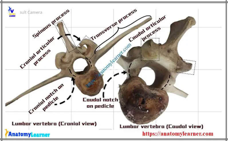
Animal lumbar vertebrae anatomy
For animal lumbar vertebrae identification, you might identify the long transverse processes from the lumbar vertebrae. The long transverse processes are the important osteological features to differentiate lumbar vertebrae from other vertebrae.
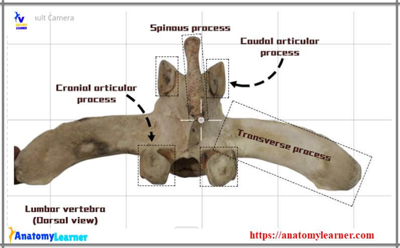
Let’s find out the following osteological features from the animal lumbar vertebrae anatomy –
#1. Presence of semi-elliptical (on cross-section) body that presents distinct ventral crest.
#2. The ventral crest diminishes from the fourth lumbar to other vertebrae
#3. Arch are equal in size (first three) as last thoracic vertebrae
#4. A caudal notch on the pedicle is much deeper than the cranial notch in the lumbar vertebrae.
#5. You will find well-developed cranial articular processes and caudal articular processes in lumbar vertebrae
#6. Transverse processes are elongated and flattened dorsoventrally, length increase in third or fourth lumbar then, diminishes.
#7. Spinous processes are in quadrilateral plates, equal in length and width diminish in the last three lumbar vertebrae.
Animal sacrum vertebrae identification
The sacrum is a single bone made with the fusion of five sacral vertebrae in cow and horse (in dog – three sacral vertebrae). It is triangular in outline and forms the roof of the pelvic cavity. This sacrum bone contains two surfaces, two borders, base and apex.
I will show you the following osteological features from the animal sacrum vertebrae identification with labeled images.
#1. Sacral spine (in a horse) or median sacral crest (cow, sheep, goat)
#2. Lateral sacral crest (low height, formed by the fusion of articular processes and parallel to the median sacral crest)
#3. Four dorsal sacral foramina (two cranially at medial aspect; two caudally – lateral to lateral sacral crest)
#4. The concave ventral or pelvic surface of sacrum bone of animals
#5. Longitudinal median groove (one) and transverse groove (four in numbers) on ventral surface of the sacrum bone
#6. Four pair of pelvic sacral foramina at ventral aspect of sacrum bone
#7. Rough borders of sacrum bone (thick portion cranially; thin portion caudally)
#8. Very wide and cranially directed base of sacrum vertebrae
#9. Wings of the base of sacrum vertebrae of animals
#10. Caudal aspect of last sacral segments (apex of sacrum bone)
#11. Sacral tuberosities
If you want to know more about sacrum vertebrae, then read the full article from anatomy learner.
Animal caudal vertebrae anatomy
These caudal vertebrae are incomplete in animals. They vary considerably in numbers in different animals. Caudal vertebrae gradually become reduced in size from the first to last vertebrae. The first three caudal vertebrae have a body, neural rings and processes. The body of the first caudal vertebrae is flattened dorsoventrally and constricted at the middle of the body.
There is a median groove (sulcus vasculosus) on the ventral surface of caudal vertebrae of animals. This median groove posses the median caudal or coccygeal artery in animals. The arch of the caudal vertebrae is small and triangular. The two flat plate forms them in animals.
Animal vertebrae identification images and videos
You have already got images and videos for animal vertebrae identification. If you need more images and videos related to animal vertebrae then, join with anatomy learner on social media for updated labeled images or videos.
Again, you might read other articles from anatomy learner like –
#1. Osteological features of hip bones of animals with labeled images
#2. Goat muscles anatomy – list of all muscles of goat body
#3. Ruminant stomach anatomy with labeled images.
Conclusion
I think this article is helpful to learn animal vertebrae identification with labeled images and videos. If you want to learn animal bone identification, read these articles related to animals’ bones here from anatomy learners.
Don’t forget to share this article with your friend who wants to know how to identify animal vertebrae or identify an animal bone. Thank you, and wait for the next article at anatomy learner.