
Hello dear anatomy learner, do you want to identify spinal cord histology slide under light microscope? Great, in this article I am going to discuss on the histological structure of spinal cord along with their identification points.
I will show you the most important histological structures from spinal cord histology slide with real images and labeled diagram. Again, you will get spinal cord histology drawing images at the end of this article.
I hope you know the spinal cord anatomy in details from different animals. If you want to memorize the anatomical features of spinal cord of cow then you might read the article from anatomy learner.
Okay, let’s get into the main part of today’s article – spinal cord histology with identification points and labeled diagram.
Spinal cord histology
You know spinal cord is the elongated and cylindrical caudal part of the central nervous system of animal. In the spinal cord histology, you will find the inner core of gray matter and outer covering of white matter. I will also discuss on these gray matter and white matter of spinal cord structure later in this article.
First I would like to enlist the important structures that you might identify from the spinal cord histology slide under light microscope at laboratory. Please try to find out these histological features from spinal cord slide images.
#1. Inner core gray matter of spinal cord (H shaped in cross section)
#2. Outer covering white matter of spinal cord
#3. Central canal of spinal cord structure
#4. Dorsal median septum of spinal cord structure
#5. Ventral median fissure of spinal cord
#6. The anterior horns of gray matter of spinal cord structure
#7. Posterior horns of gray matter of spinal structure
#8. Axon fibers on the white matter of spinal cord
#9. Motor neuron at gray matter of spinal cord
#10. Dura matter, pia matter and arachnoid matter surrounds spinal cord (if possible)
#11. Fasciculus at white matter
#12. Gray commissure of gray matter
Okay, I tried to identify most of these structures at spinal cord histology slide images.
Spinal cord histology identification points from slide images
Okay, now I am going to provide you the best and important identifying characteristics of spinal cord under light microscope. If possible you may enlist more spinal cord histology identification points.
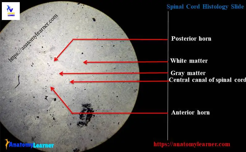
#1. Presence of dark brown outer white matter and the light staining inner gray matter in the cross section of spinal cord
#2. There is H shaped gray matter (two parts at two lateral sides which connected each other at midline of spinal cord by gray commissure)
#3. Presence of central canal at the center of the gray commissure of spinal cord structure
#4. The central canal is line by the ependymal cell (tall columnar cell)
#5. Presence of cell bodies of neurons and interneurons at the gray matter of spinal cord structure
#6. There are ascending and descending myelinated nerve fibers present at the white matter of spinal cord structure
#7. The anterior horns of gray matter are more prominent than the posterior horns of gray matter
So these histological features confirm that this is a slide of spinal cord.
Spinal cord histology slide with labeled diagram
Do you want to learn details histology of spinal cord with real slide images and labeled diagram? Fine, you might continue this article to learn more about the histological features of spinal cord.
You know there are two main parts of spinal cord structure – inner core gray matter with central canal (lightly stained) and outer darkly stained white matter (surrounds the gray matter). Let’s know about the histological features of gray matter and white matter of spinal cord.
Histological features of gray matter of spinal-cord
In gray matter histology you will find the H shaped structure with anterior and posterior horns which is connected by a gray commissure. In this gray commissure you will find the central canal that is lined by ependymal cell.
The gray matter of spinal cord consists of the following structures –
#1. Nerve cell bodies and their processes
#2. Neuroglia cells and
#3. Numerous blood vessels
You will find large multipolar neurons cell bodies in the anterior horns of gray matter. You know the anterior horn of gray matter extends toward the front of the spinal cord and are more prominent than the posterior horn. Again, the posterior horns are the sensory area and contain the smaller neurons cells bodies.
White matter of spinal-cord structure
Gray matter of spinal cord is surrounded by the white matter. Entire white matter divides into two lateral halves with anterior median fissure and ventral median septum.
In white matter of spinal cord histology you will find the following important structures –
#1. Different nerve fibers
#2. Neuroglia cells at white matter of spinal cord and
#3. More blood vessels at white matter of spinal cord structure
The spinal cord is surrounded by connective tissue meninges, consisting of outer dura matter, middle arachnoid matter and inner pia matter.
How to differentiate different segments of spinal cord structure?
You may differentiate the different segments of spinal cord structure under light microscope. In cervical segment of spinal cord structure you will find the increased gray matter particularly at the anterior horns. This occurs because of large collection of the nerve cell bodies in gray matter.
Again in thoracic segment of spinal cord structure you will find the small gray matter, the anterior and posterior horns are narrow. In lumbar and sacral segment of gray matter you will find the increased gray matter.
Spinal cord histology drawing
I am going to show you the spinal cord histology drawing for better understanding. Here I tried to draw all of the important structures from gray and white matter of spinal cord histology. You may follow this drawing but try to make more appropriate.
If you need more drawing and real images related to spinal cord slide then you may follow anatomy learner at here in social media (for latest updated pictures).
If you want you might like other article from anatomy learner (nervous tissue and system of animals) –
#1. Histological features of cerebellum with identifying points and
#2. Identifying features of cerebrum with real slide picture
Conclusion
I know you got the best guide to learn spinal cord histology with real slide images and labeled diagram. Now you will able to identify the spinal cord slide with identification points that I mentioned in this article.
If you think this article is helpful to learn gray matter and white matter structures of spinal cord then you may share this with your friends who want to learn histological features of spinal cord.
Don’t forget to follow anatomy learner’s social media and check the latest articles from anatomy learner blog