
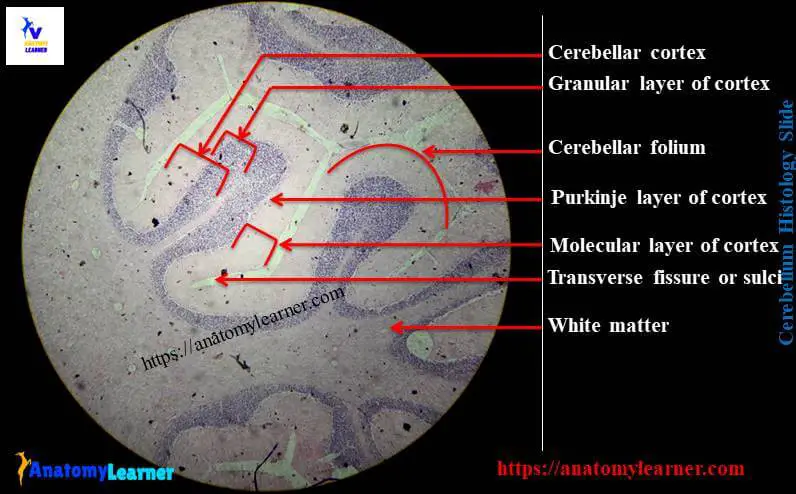
Cerebellum histology consists of outer gray matter and inner white matter in different animals. The outer gray matter of cerebellum found mainly the covering surface and known as cortex of cerebellum. On the other hand, white matter found in the central part of cerebellum and known as medulla of cerebellum.
Hey, do you looking for the best guide to learn cerebellum histology with real slide labeled diagram? In this article I am going to share the cerebellum histology layers along with the identification points under light microscope.
So you have taken your good decision to getting into this cerebellar histology article. In this article you will learn the histology of cerebellar cortex and cerebellar white matter from cerebellum.
After completing this article you will able to understand the different layers and cells from cerebellum histology slide labeled diagram. I will also share cerebellum histology drawing and ppt with you so that you might practice.
So, if you are interest to learn cerebellum histology then I would like to request you to read this complete guide. Hope this guide will help you to learn the basic of cerebellar cortex histology.
You may learn cerebrum histology slide with labeled diagram from this anatomy learner blog. Okay, let’s get into the main part of this article – histological features of cerebellum cortex and medulla in animal.
Cerebellum histology
From cerebellum histology, I will show you the following important histological features under light microscope –
#1. Narrow ridge or folia at cerebellum surface
#2. Transverse groove or sulci at cerebellum surface
#3. Molecular layer of cerebellum cortex in animal
#4. Individual purkinje cell and purkinje cells layer in cerebellar cortex
#5. Dendrite of piriform cells in cerebellar cortex
#6.Granular cells layer in cerebral cortex in animal
Let’s find these structures and cells from the cerebellum cortex histology labeled diagram. Hope you could understand the histological structures of animal cerebellum.
Cerebellum histology identification points from slide
Do you want to identify the cerebellum histology slide under light microscope at laboratory? Fine, here I am going to enlist the most important cerebellum histology identification points. You could identify cerebellar slide with following identifying characteristics.
#1. Presence of highly folded surface at cerebellum (also known as folia) which is separated by transverse groove or sulci
#2. Thick and light stained molecular layer at the superficial part of cerebellar cortex
#3. Presence of single layer of large flasked shaped (larger cell body) purkinje cell layer at deep to the molecular layer of cerebellar cortex
#4. There are densely packed granular cells (small neuron with heterochromatic nuclei) at deep to the purkinje cell layer and close to white matter of cerebellum
These are the main identifying characteristics of cerebellar histology slide under light microscope. You may add other identifying characteristics to identify cerebellum under light microscope.
Cerebellum histology layers with labeled diagram
Do you want to know more about the cerebellum histology layers? You know cerebellum consists of outer gray matter (cortex) and inner white matter (medulla) in animal. I am going to discuss on cerebellar cortex histology and cerebellar white matter histology separately. Let’s start to know the histological features from these two parts of cerebellum of animal –
#1. Histology of cerebellar cortex in animal and
#2. Histology of cerebellar white matter in animal
Cerebellar cortex histology
You will find the well-defined three layers in cerebellar cortex histology in animal. The cerebellar cortex is located at the periphery of the cerebellum.
The cortex of cerebellar part is highly folded and these folded parts are known as folia (narrow ridge). These folia are separated by the transverse groove and these transverse grooves are known as sulci or transverse fissure.
Each of this folium contains a center white mater which is surrounded by gray matter superficially.
You will find the following layers at the cerebellar cortex in animal –
#1. Outer molecular layer of cerebellar cortex in animal
#2. Middle purkinje cell layer of cerebellar cortex and
#3. Inner granular layer of cerebral cortex
Outer molecular layer from cerebellum histology
You will find the following histological features in outer molecular layer of cerebellar cortex in animal.
#1. This outer molecular layer is thick and located superficially at cerebellum
#2. It is light stained and you will find four important features – stellate cells, basket cells, dendrite of purkinje cells and unmyelinated axons from granular cells.
#3. The stellate cells located at the superficial layer of cerebellar cortex and basket cell locates deep to stellate cells
#4. The axon of basket cell courses the folium transversely and surrounds purkinje or piriform neuron cells body
Middle granular layer
In middle granular cells layer, you will find the following important histological features.
This purkinje cell layer contains larger flask shaped cell body purkinje cell or piriform cells. These piriform cells are arranged in a single row in between the molecular and granular layer of cerebellar cortex.
The axon of purkinje cells goes to the white matter of cerebellum and synapse with neuron of cerebral nuclei. Again the purkinje cell has elaborative dendrite that projected into the molecular layer.
Inner granular layer of cerebral cortex
In cerebellum histology, you will find densely packed granular cells located adjacent to the white matter of cerebellum. These cells have small body with heterochromatic nuclei at cerebral cortex of animal brain.
The axon of the granular cells enter the molecular layer and bifurcates, travel longitudinally within the folium of cerebral cortex. These axons of granular cells synapse with the dendrite of the piriform or purkinje cells.
There are two types of major nerve fiber enter into the cerebral cortex of animal – climbing fibers and mossy fibers.
The climbing fiber climbs like vines and synapse with the dendrite of purkinje cells. Again the mossy fiber has expanded terminal part and synapse with granular cells.
Cerebellum histology drawing
Do you need the cerebellum histology drawing? Fine, here I am going to share the drawing of cerebellar histology. You may follow the same cerebellar histology drawing and try to perform better. Please focus on the every single structures and layers of cerebellar cortex and cerebrum medulla in your drawing section
Cerebellar white matter or medulla
White matter is the center part of the cerebellum of animal brain. Histologically the cerebellar white matter of cerebellum consists of myelinated nerve fibers.
Cerebrum histology slide labeled diagram
If you want to know more about cerebrum histology slide with labeled diagram then you might find this article here in this anatomy learner blog. Let’s find the different layers of cerebrum histology slide from the labeled picture below –
You might learn the other histological features from anatomy learner blog – like spinal cord histology, structure of a neuron or other.
If you need more pictures related to cerebellum histology then please follow anatomy learner at here in social media.
Conclusion
Hope you could able to learn the cerebellum histology of animal with labeled diagram. If you think this guide was helpful for you and you really learn about cerebellar histology, then share this article with your friends who want to learn cerebellum histology.
Again, if you need cerebellum histology ppt for practice then let me inform. Read other article – cerebrum histology with labeled diagram from anatomy learner blog.