
You know, gland is an organ of secretion made of specialized secretory cells that is derived from the surface epithelium on which it opens. There are different types of gland like exocrine, endocrine, serous and others in the animal body. These glands of animal body are classified based on different criteria. In this article I will discuss on the salivary gland histology with their important identification points.
There are three major salivary gland in animal body – parotid, submandibular and sublingual salivary glands. Parotid gland is compound acinar gland whereas submandibular and sublingual glands are compound tubuloacinar in nature.
Here, I am going to discuss the basic histology of salivary glands (parotid, submandibular and sublingual gland) with real slide pictures and also with labeled diagram.
I will also discuss on the basic structure of salivary gland histology and the structure of mucous and serous acini. Again at the end of this article you will get the salivary gland histology drawing.
So, do you love to learn the basic histology of salivary glands with their identifying characteristics? Fine, let’s get into the article – salivary gland histology with real slide images and labeled diagram.
Salivary gland histology
Before going to details salivary gland histology, I would like to provide the following information for your better understand. If you have good ideas on these topics then you might skips –
#1. General structure of a compound gland like salivary gland
#2. Serous acini structure
#3. Mucous acini structure and
#4. Structure of serous demilune
Okay, let’s start to learn about them little.
Structure of a salivary or compound gland
In compound gland you will find the serous or mucous or both types of acini or secretory cells. These cells are often associated with the contractile myoepithelial cells. There is a dense or loose connective tissue framework surrounds the gland and supports the parenchyma and forms the stroma of the gland.
In the parenchyma of compound gland you will find the secretory unit and it may be acini or tubules or tubule-acini. Again, you will find the different ducts like interlobular, interlobular or execratory ducts on parenchyma of compound gland structure. In stroma you will find the loose connective tissue supporting framework along with capsule and septa.
Serous acini structure
Serous acini secrete the thin watery secretion and rich with enzymes. This acinus has pyramidal shaped cells with round, centrally located nucleus. The lumen of the serous acini is narrow compare to the the lumen of mucous acini. These serous acini stained darkly in routine staining. In parotid salivary gland histology slide, you might find the serous acini. All serous acini are surrounded by a thin and contractile myoepithelial cells.
Structure of mucous acini
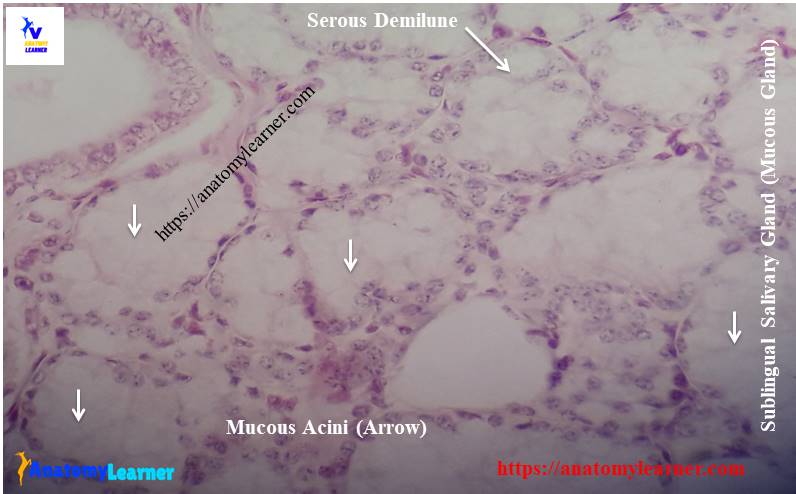
Mucous acini secrete the thick viscous materials and contain mucigen droplets on cell surface. They have flattened and peripherally placed nucleus and large lumen. In routine stain, they take the light stain; you might find the mucous acini in sublingual salivary gland histology slide.
Seromucous or serous demilune structure
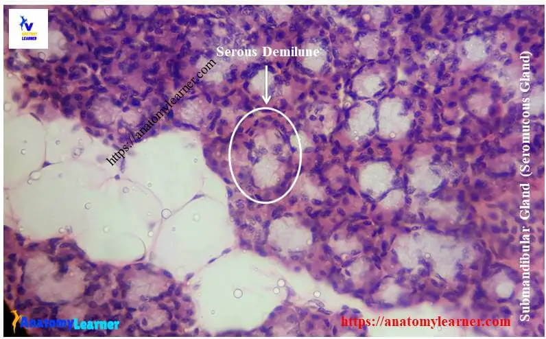
You will find both the serous acini and mucous acini in this acinus. In submandibular salivary gland histology slide you will find the serous demilune where the mucous acinus is surrounded by serous acinus at one side.
Fine, now you will able to identify the different salivary gland structure under light microscope.
Parotid salivary gland histology with labeled diagram
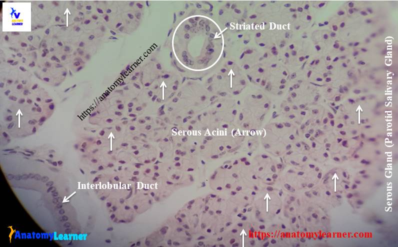
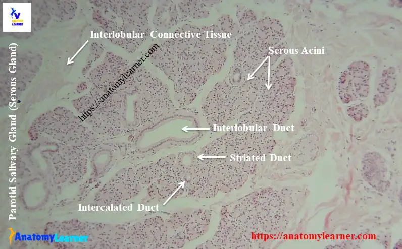
Parotid salivary gland is the large serous gland which is surrounded by a thick connective tissue capsule. This capsule extends into the parotid gland and subdivides the gland into numerous lobes and lobules. In each lobule you will find the darkly stained serous acini cells. You will also find the ducts system (striated ducts, intralobular excreatory ducts, intercalated ducts) at parenchyma of parotid gland.
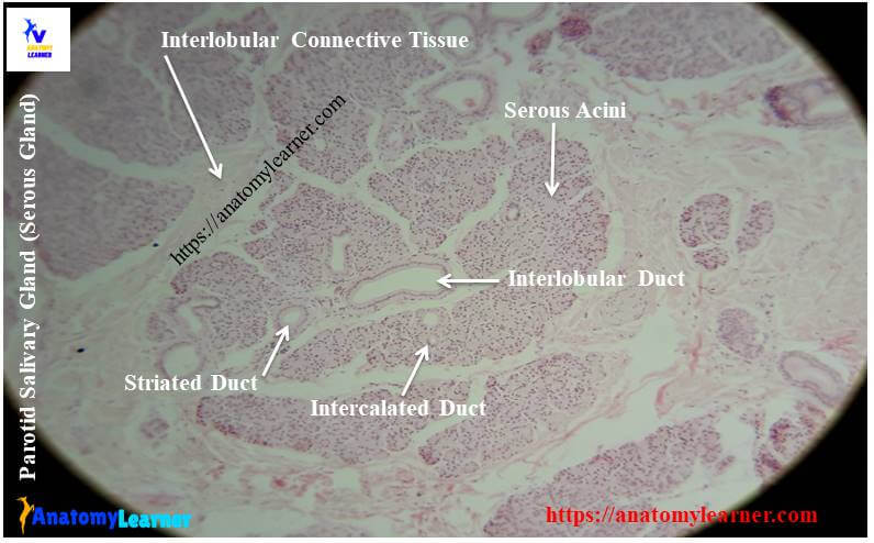
Secretory products from serous acini empty into a narrow channel termed as intercalated ducts. The lumens of these intercalated ducts are larger and lined by simple squamous or low cuboidal epithelium. The intercalated ducts drain the secretory materials into a large striated duct which have a larger lumen. The luminal surface of striated duct is lined by simple columnar epithelium. Again the striated ducts empty their materials into the intralobular execratory duct of parotid gland. These intralobular execratory ducts of parotid gland are lined by simple cuboidal epithelium.
From the parotid salivary gland histology slide you might identify the following structures under light microscope –
#1. Serous acini in parotid gland structure
#2. Striated ducts on parenchyma of parotid gland
#3. Intercalated duct of parotid gland structure
#4. Interlobular ducts on parotid parenchyma
#5. Interlobular connective tissue septum of parotid gland structure
Identifying characteristics of parotid salivary gland slide
Do you want to identify the parotid salivary gland histology slide under light microscope? Well, following identifying characteristics of parotid salivary structure will help you to identify parotid slide under light microscope.
#1. Presence of darkly stained serous acini with narrow lumen in the section
#2. Nuclei is round and centrally placed in the serous acini cell
#3. Intercalated duct, striated ducts, intralobular and interlobular ducts are well developed in the parotid gland section
So, this is a slide of parotid salivary gland of animal.
Submandibular salivary gland histology
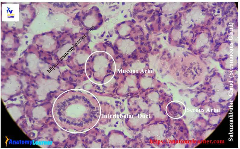
You know, submandibular salivary gland is a compound tubuloacinar gland in animal. This gland is a mixed gland that contains both serous acini and mucous acini. You will also find the same structure in the stroma and the parenchyma of submandibular salivary gland histology. This submandibular salivary gland is covered by a connective tissue capsule and extends into the gland parenchyma and divided the gland into different lobes and lobules.
In each lobules of submandibular salivary gland contain the darkly stained serous acini cells and lightly stained mucous acini cells. The mucous acini of submandibular gland are normally capped by one or more serous cells and forms a cresent shaped serous demilune. The duct system are poorly developed
You might identify the following important features from the submandibular salivary gland histology slide under light microscope –
#1. Serous and mucous acinus in the submandibular gland slide
#2. Serous demilune in the section of submandibular gland
#3. Interlobular and striated ducts of submandibular gland
#4. Connective tissue capsule and septa of submandibular gland
Identification points of submandibular salivary gland slide
If you want to identify submandibular salivary gland slide under light microscope then the following important identification points might help you.
#1. Presence of both darkly stained serous acinus and lightly stained mucous acinus in the section
#2. Cresent shaped serous demilune is present (mucous acini caps with one or more serous acini)
#3. Presence of moderately developed duct system in the gland parenchyma
So this is a submandibular salivary gland histology slide. You may add other different identifying characteristics of submandibular gland structure.
Sublingual gland histology
The sublingual salivary gland is a mixed and compound tubuloacinar gland that contains both the serous and mucous acini. But you will find more numbers of mucous acini than the serous acini in any section of the gland. Actually pure serous acini are less in number in the histology of sublingual salivary gland histology.
Most of the mucous acini cells are capped with serous acini which are actually known as serous demilune. Both the serous and mucous acini of sublingual salivary gland are surrounded by the contractile myoepithelial cells.
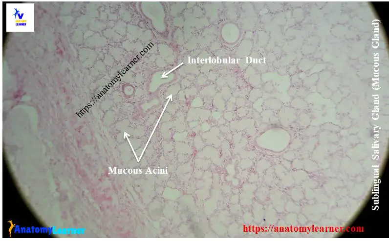
The duct system of sublingual salivary gland is somewhat different from other salivary gland like parotid or submandibular gland. Intercalated ducts are short or absent and the nonstriated interlobular excreatory ducts are more in numbers in sublingual salivary gland histology section.
From the sublingual salivary gland slide you might also identify the following histological features –
#1. More mucous acini and less number of serous acini
#2. Serous demilune of sublingual gland structure
#3. Interlobular and striated ducts of sublingual gland
#4. Interlobular septum of sublingual lobules
Identifying characteristics of sublingual gland slide
Okay, now it is time to identify the sublingual salivary gland histology slide under light microscope. Hope the following identifying characteristics will help you to identify sublingual salivary gland slide.
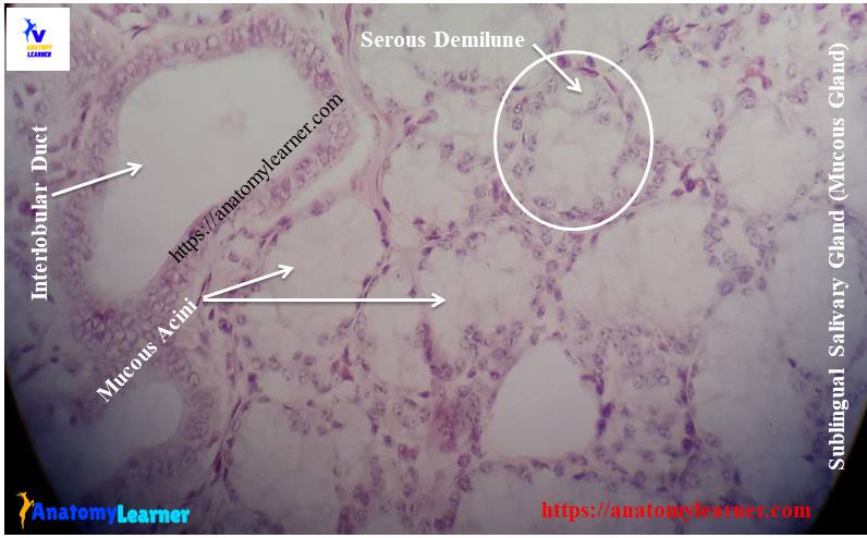
#1. Presence of more numbers of lightly stained mucous acini or tubules with large lumen in the section
#2. The nuclei of the mucous acini is flat and located at the periphery of the cell
#3. Presence of poorly developed duct system in the section of this gland
So, this is the slide of sublingual salivary gland.
Salivary gland histology drawing
Now I am going to provide salivary gland histology drawing for better understand. You might try to draw the better images then me.
If you need more images related to salivary gland structure then you may follow anatomy learner at social media (get more pictures of salivary gland).
Again you might read other different article from anatomy learner blog (get the latest article at here).
#1. Histological features of mammary gland with real images and labeled diagram
#2. Identification of pituitary gland under light microscope with important identifying characteristics
Conclusion
I hope you learn the basic of salivary gland histology from this article. Now you will able to identify the salivary gland slide under light microscope.
If you find any mistake in salivary gland histology labeled diagram or slide images then please let me inform. Again, if you need more parotid gland histology diagram or slide pictures then you may follow anatomy learner at social media.
Do this article helpful for you to learn salivary gland histology with their identifying characteristics? If you found this article was helpful to learn salivary gland structure then share it with your friend who also want to learn the same.
Thank you so much for staying with anatomy learner and learn veterinary gross anatomy and veterinary histology with me.