
Spleen is the major secondary lymphatic organ in animal involved in filtrating blood and mounting immune response. Normally spleen is a blood forming organ in foetal life and blood destroying organ in postnatal life. In spleen histology, you will find the two important structures – connective tisuue framework and parenchyma.
Welcome back to anatomy learner and thanks for getting into this article – spleen histology red and white pulp with slide images and labeled diagram. If your are looking for the best guide to learn histological features of spleen structure then you may continue this article.
I am going to discuss on the basic histology of spleen along with identification points under light microscope. After completing this article you will able to identify the spleen histology slide under light microscope based on their specific identifying characteristics.
Here I will show you the different structures from white and red pulp of spleen structure under light microscope with real slide images. You will also get the spleen histology drawing for better understand at the end of this article.
Spleen histology
In the parenchyma of spleen histology slide, you will find the red pulp and white pulp. Before going to details description of spleen structure I would like to enlist the important structures that you might identify under light microscope.
Fine, here are some important structures of spleen histology slide –
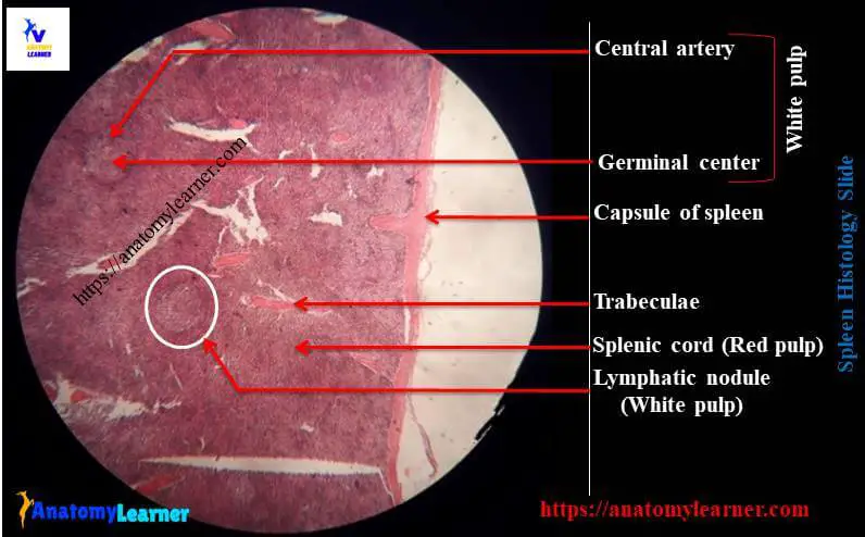
#1. Thick capsule and trabeculae of spleen
#2. White pulp area in the section of spleen
#3. Germinal center of white pulp area
#4. Central arteriole in the white pulp of spleen structure
#5. Red pulp of spleen histology
#6. Spleenic cords in red pulp of spleen
#7. Venous sinus in red pulp of spleen
Well, let’s try to find out these structures from the spleen slide.
Spleen histology identification
Do you want to identify the spleen slide under light microscope? Fine, I am going to enlist some spleen histology slide identification points that might help you to identify spleen. Here I am going to share the most important and specific identifying characteristics of spleen structure, but you may enlist other points also.
#1. The tissue section surrounds by a thick dense connective tissue capsule from which the trabeculae extends deep into the structure
#2. Presence of aggregations of lymphatic nodules in the section termed as white pulp
#3. Each ymphatic nodules traversed by a blood vessel called central arteriole
#4. Diffuse cellular networks present around the lymphatic nodules termed as red pulp
#5. Presence of splenic cords and sinusoids in the red pulp of the tissue section
So, this is a spleen histology slide.
Spleen histology red white pulp with labeled diagram
So, do you want to learn the details histology of spleen with real slide images and labeled diagram? Well, I will guide you to learn the details histological structure of spleen in easy way.
You know that there are two important components in spleen structure – one is connective tissue framework and another parenchyma. Again, you will find the following structures under connective tissue framework and parenchyma of spleen.
In the connective tissue framework of spleen, you will find the followings –
#1. Thick dense connective tissue capsule
#2. Thick connective tissue trabeculae of spleen
#3. Reticular stroma of spleen
In the parenchyma of spleen you will find the following main structures –
#1. Red pulp of spleen and
#2. White pulp of spleen (large aggregation of lymphoid tissue)
Okay, now let’s discuss on the framework and parenchyma of spleen separately.
Connective tissue framework of spleen
The spleen is surrounded by thick dense connective tissue capsule and it consists of two layers – layer of dense irregular connective tissue and a layer of smooth muscle. The thickness and relative amount of smooth muscle in the capsule may vary with different animals.
From the capsule, trabeculae extends deep into the spleen’s interior. These trabecuale of spleen are thick and robust. Trabeculae mainly composed of collagen fibers, elastic fibers and smooth muscle cells and also contains arteries, veins, lymph vessels and nerves. You will also find the reticular fibers and phagocytic reticular cells throughout the spleen.
All these capsule, trabeculae and reticular fibers supports the splenic parenchyma.
Parenchyma of spleen structure
The parenchyma of spleen histology composed of red pulp and white pulp. Do you want to know the histological features of red and white pulp of spleen? Fine, let’s continue this article to know more about the white pulp and red pulp of spleen structure.
White pulp of spleen
The white pulp of spleen structure is lymphatic tissue that is distributed throughtout the spleen. It comprises of lymphatic nodules and diffuse lymphatic tissue called periarterial lymphatic sheaths.
So the white pulp of spleen is mainly formed by the numerous aggregations of lymphatic nodules (lymphocytes and lymphoblasts). You will find germinal center with large collection of B lymphocytes and central arteriole within the lymphatic nodules of white pulp of spleen.
The lymphatic nodules is surrounded by the an immunologically active zone containing macrophages, few T lymphocytes and blood vessels. The functional zone between the white and red pulp is known as the marginal zone or area of spleen.
The central artery of white pulp area locates at the periphery of the lymphatic nodule.
Red pulp of spleen structure
The red pulp is the modified lymphoid tissue that is infiltrated with all the cells of circulating blood. This gives the dark red color to the spleen tissue in fresh condition.
There are two main components in the red pulp of spleen histology – venous sinuses or venules and splenic cords.
The splenic sinuses are wide vascular channels that lines with elongated, longitudinally oriented endothelial cells. The narrow splenic cords located between the sinuses and forms a three dimensional network. This cord contains the reticular fibers, reticular cells, erythrocytes, macrophages, lymphocytes, plasma cells and other leukocytes.
In the red pulp of ruminant and pig spleen, you will find numerous smooth muscle cells but in horse spleen you will find the myofibroblast in red pulp.
Spleen histology drawing
Okay, now I am going to share spleen histology drawing with you so that you might draw the same images. But make sure you try to draw the better spleen slide image than me.
If you need more spleen drawing images or real slide images or labeled diagram then you may follow anatomy learner in social media (for more spleen slide images).
Conclusion
I hope you learn the basic of spleen histology with slide images and labeled diagram. Now you could able to identify the histological features of spleen under light microscope.
If you think this article is helpful to learn spleen structure then you may share this with your friend who also wants to learn histology of spleen.
Thank you so much for staying with anatomy learner and don’t forget to check the latest article from anatomy learner.