Tonsil Histology Slide with Labeled Diagram – Histological Features of Palatine and Lingual tonsils

Tonsil histology slide microscope image

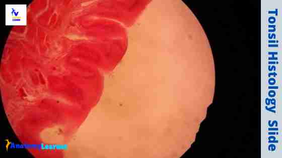
Near the junction of the oral cavity and the pharynx, you will find several collections of lymphoid tissue that refer to the tonsil. In tonsil histology, different essential structures like tonsillar crypts, lining epithelium, numerous lymphatic nodules, and dense connective tissue capsules should be identified under a light microscope.  I will show you all these structures from … Read more

Thymus Histology – Normal Features Identification and Real Slide Pictures

Thymus histology

Thymus of animal is a central lymphoid organ which is essential for the growth and development of other lymphoid organs. In thymus histology you will find the dark staining outer cortex and light staining inner medulla.  Hi, do you want to identify normal thymus histology slide under the light microscope with proper identifying characteristics? In … Read more

Spleen Histology – White Pulp and Red Pulp Histology with Labeled Diagram

Spleen histology

Spleen is the major secondary lymphatic organ in animal involved in filtrating blood and mounting immune response. Normally spleen is a blood forming organ in foetal life and blood destroying organ in postnatal life. In spleen histology, you will find the two important structures – connective tisuue framework and parenchyma. Welcome back to anatomy learner … Read more

Lymph Node Histology – Cortex and Medulla Description

Lymph node histology

Lymph node is a oval or bean shaped structure located along the extensive drainage system of lymph vessels of animal. In lymph node histology, you will also find two main components – the connective tissue thin framework and parenchyma. Hi there, welcome back to anatomy learner and thank you so much for getting into this … Read more