The height of the simple cuboidal epithelium is about the same as its width. Here, I will answer the question – ‘Where is simple cuboidal epithelium found?’
Quick answer: simple cuboidal epithelium can be found in the lining of the thyroid follicles, kidney tubules, and on the surface of the ovary. It is also present in the brain’s choroid plexus, the eye’s ciliary body, and retinal pigment.
I will show the simple cuboidal epithelium from this organ under the light microscope. Again, I will provide identifying features of the simple cuboidal epithelium.
Where is simple cuboidal epithelium found?
The simple cuboidal cells are arranged in a single layer resting on the basement membrane. These cells are cubical and have central rounded nuclei.
This simple cuboidal epithelium may be found in the ducts and follicles of most of the glands. Here, Table 1 shows the location of the simple cuboidal epithelium from different organs –
| Simple cuboidal epithelium location | Specific parts of the organs |
| Thyroid glands | Ducts and follicles of thyroids |
| Ovary | The outer surface of the animal’s ovary |
| Kidney tubules | Proximal convoluted tubules of the kidney Collecting ducts of the kidney |
| Brain | Choroid plexus of the brain |
| Eye | The ciliary body of the animal eye The inner surface of the lens Pigment cell layers of the retina |
So, the epithelium of the thyroid, ovary, kidney, brain, and specific parts of the eye are simply cuboidal.
What does the simple cuboidal epithelium look like under the light microscope?
The simple cuboidal cells appear as squares in the cross-section under the light microscope. But they look hexagonal-shaped when seen from the surface.
The simple cuboidal epithelium is the single layer of cells that possess the same height and width. Thus, they are known as the simple cuboidal epithelium.
But, when the height is slightly less than the width of the cells, it is known as the low cuboidal epithelium. When the height is slightly greater than the width, this epithelium is known as the tall cuboidal epithelium.
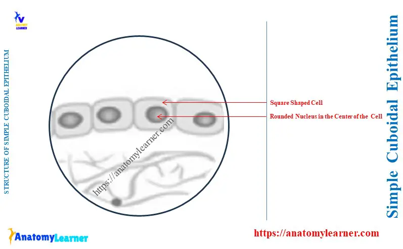
The nuclei of the simple cuboidal epithelium are rounded. It is located at the center of the simple cuboidal epithelium.
Here, the diagram shows the square-shaped cells and rounded nuclei of the simple cuboidal epithelium. The diagram also shows the rounded nucleus is located at the center of the square-shaped cells.
Identifying features of the simple cuboidal epithelium
The below-mentioned identifying features might help you to identify the simple cuboidal under the light microscope –
- The epithelium cells are arranged in a single layer that rests on the basement membrane,
- Presence of square-shaped cells (in cross-section) that have the same length, breadth, and height and
- The presence of a rounded nucleus in the center of the epithelial cells,
So, the shape of the cells and nucleus and their arrangement are the key identifying features of simple cuboidal epithelium.
Is simple cuboidal epithelium found in the glands?
Answer: Yes, the simple cuboidal epithelium may be found in the glands. They are typically located in the lining of the follicles and ducts of the glands.
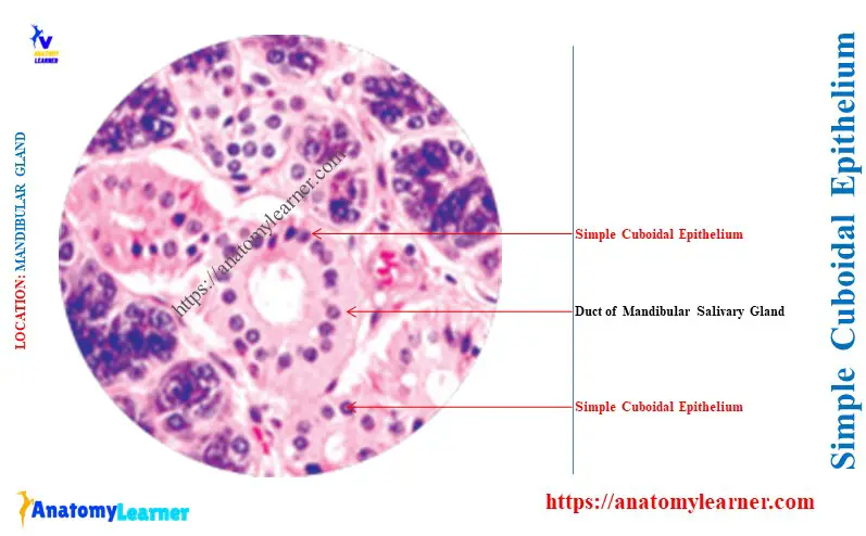
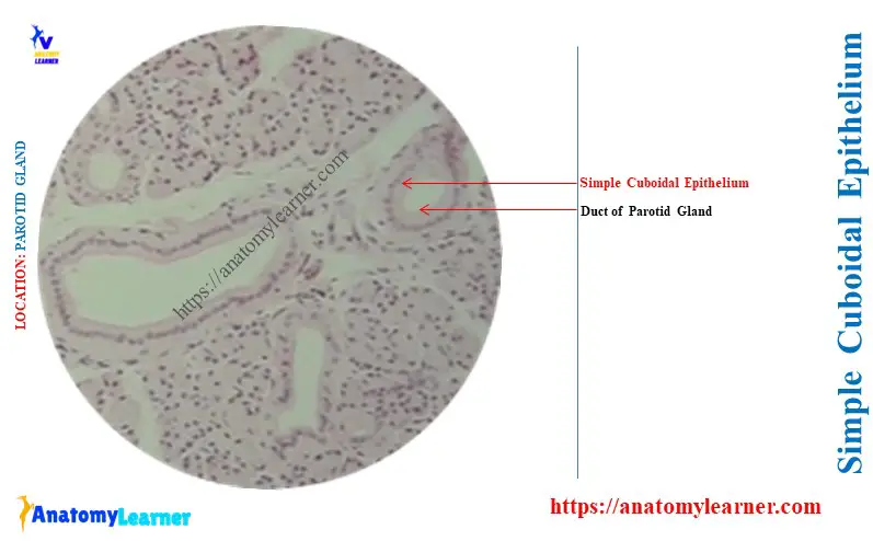
For example, the follicles of the thyroid and parathyroid glands are lined by the simple cuboidal epithelium. Again, the ducts of the thyroid and major salivary glands have the lining of the simple cuboidal epithelium.
Here, the diagram shows the simple cuboidal epithelium from the ducts of the parotid and submandibular salivary glands. But, you may find the stratified cuboidal epithelium lining in the excretory intralobular ducts of these salivary glands.
Is simple cuboidal epithelium found in the thyroid?
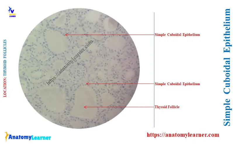
Answer: yes, the simple cuboidal epithelium may be found in the thyroid’s follicles and ducts. Here, the diagram shows the simple cuboidal epithelium from the follicles of the thyroid glands.
The epithelium of the thyroid follicles is made up of cells that look like a square. It also presents the centrally placed rounded nuclei in the thyroid follicles.
Simple cuboidal epithelium in ovary and kidney tubules
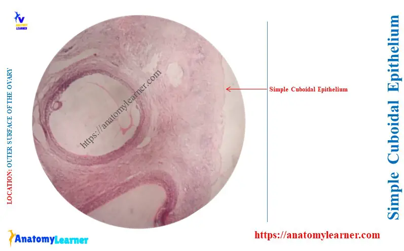
The surface of the animal ovary lines with a single layer of simple cuboidal epithelium. This type of epithelium of the ovary is the germinal epithelium.
Here, the diagram shows the simple cuboidal epithelium from the outer surface of the ovary. The microscopic features of this germinal epithelium are similar to the typical simple cuboidal epithelium.
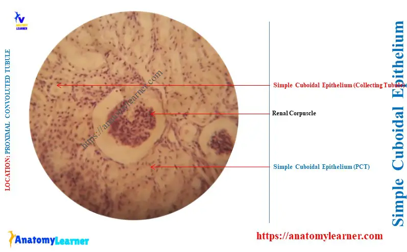
The kidney tubules, especially the proximal convoluted tubules and collecting ducts, show a simple cuboidal lining. Here, the diagram shows the simple cuboidal lining from the proximal convoluted tubules of the animal kidney.
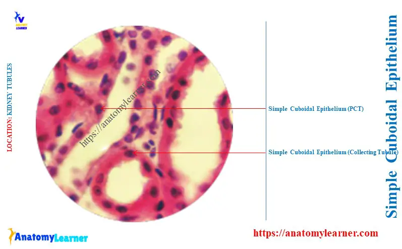
The shape, size, and staining intensity of the kidney tubules may vary. To examine the simple cuboidal from the tubule, you might observe them under high magnification.
The outline of the cuboidal cells may not be visible due to poor preservation and processing. But you may confirm the cell shape by observing the round nuclei as ring-shaped around the tubules.
You know the retina of the animal eye has 10 layers, whereas the pigment layer has special epithelium. This layer of the retina consists of simple cuboidal epithelium.
Conclusion
Thus, you got the specific answer to the question – ‘Where is simple cuboidal epithelium found?’ The ducts and follicles of most glands, kidney tubules, and outer surface of the ovary show the typical simple cuboidal epithelium lining.