
Kidney is the organ of urinary system and involves in production of urine in animal that conveyed by the ureters to the urinary bladder. In kidney histology, you will find the outer darker cortex and inner lighter staining medulla.
Hi there, welcome back again and thanks for getting into this article – kidney histology with identification point along with labeled pictures and drawing. Okay, if you are looking for the best guide to learn different histological structures of kidney from kidney slide then this article may help you a lot. In this article I am going to show you the every single structures of cortex and medulla from kidney histology slide.
You will also get a full concept on the different structures of a renal corpuscle with real slide picture. After completing this article you will able to identify the kidney slide under light microscope at laboratory. Again, you will get different kidney histology labeled drawing, diagram and pictures at the end of the article.
So, if you really want to learn the histological basis of kidney then I would like to suggest you to continue this article till end.
Kidney histology
I hope you know the details anatomy of kidney from different animals. The kindey of different domesticated animals have various shaped and sized. You know, the outer surface of the kidney is covered by a fibrous capsule; histologicall it is composed primarly by collagen fibers but also contains smooth muscles and different blood vessels.
First, I would like to enlist these structure that you might identify from the kidney histology slide under light microscope. Here I am going to enlist the different histological structures of kidney from medulla and cortex.
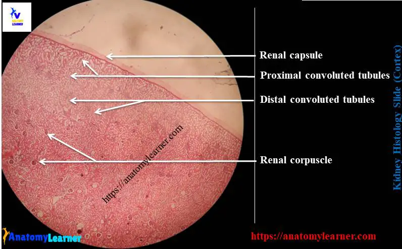
Histological structures from kidney cortex –
#1. Connective tissue capsule of kidney
#2. Proximal convulated tubules of kidney cortex
#3. Distal convulated tubules of kidney cortex
#4. Renal corpuscle of kidney cortex
From kidney medulla you might identify the following histological structres
#1. Collecting ducts and collecting tubules of kidney medulla
#2. Thin descending limb of loop of henle of kidney medulla
#3. Thick ascending limb of loop of henle of kidney
#4. Interistium and differnet capillaries at kidney medulla
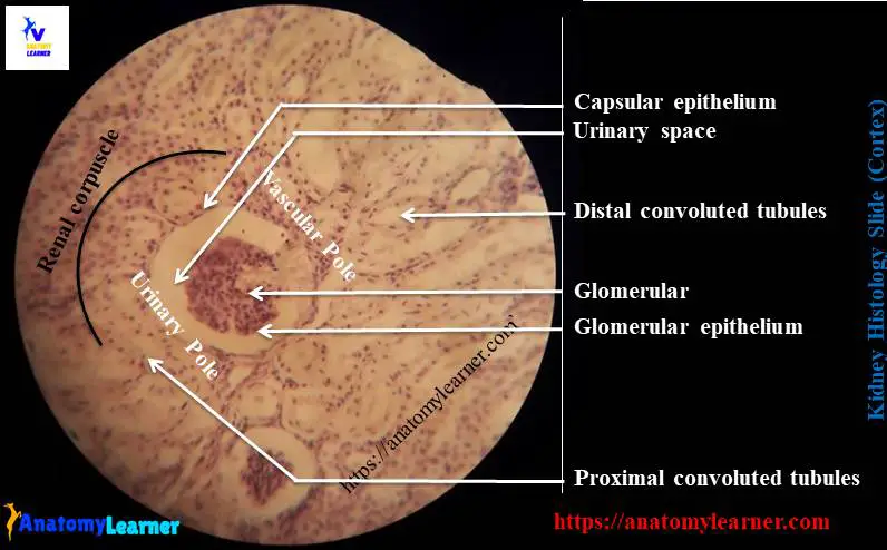
Again, form the reanl corpuscle of kidney cortex histology, you might identify the following features –
#1. Vascular pole of reanl corpuscles
#2. Urinary pole of renal corpuscle
#3. Bowman’s capsule of corpuscle
#4. Capsular epithelium
#5. Glomerulus of corpuscle
#6. Glomerulus epithelium
Identifying characteristics of kidney histology slide
You may asked to identify different structures from kidney histology slide. Then you might write their specific histological features of specific structures of kidney. I am going to enlist the identifying characteristics of overall kidney slide under light microscope.
#1. Presence of outer darker or dark red cortex and inner lighter color stained medulla in the section
#2. There is a connective tissue capsule present at the surface of organ structure (termed as renal capsule)
#3. Presence of renal corpuscle, proximal convulated tubules (darkly stained; lined by simple cuboidal epithelium with microvilli) and distal convulated tubules (lightly stained; lined by simple cuboidal but no microvilli) in the dark cortex area
#4. There are collecting ducts, collecting tubules, thick loop of henle and thick loop of helne in the inner lighter meduall area
So, this is a slide of kidney histology. You may also asked to identify only the renal corpuscle of kidney; then let’s write the following identifying characteristics –
#1. Presence of Bowman;s capsule; the parietal layeris lined by the simple squamous epithelium
#2. There are urinary space or bowman;s space present in between the parietal and visceral layers of bowmans capsule
Histological features of kidney cortex and medulla with labeled diagram
Histologically, kidney is composed of many compactely packed renal tubules that embedded in a vascular interistium. Each of these renal tubules contain two distinct starurces – nephron and collecting tubules.
You know, nephron is the staructural and functional part of kidney and mainly composed of the following parts –
#1. Renal corpuscle
#2. Proximal convulated tubules
#3. Loop of henle (thin descending and thick ascending loop) and
#4. Distal convulated tubules
Renal corpuscle structure
The renal corpuscle is composed of the glomerulus capillaries tuft (mesengium) and the glomerulus capsule (known as bowman’s capsule). The shape of renal corpuscle is sheprical but the size varies in different animals.
The glomerulus cappilaries tuft are actually is a network of branching and anastomosing capillaries which is lined by simple squamous epithelium (fenestated endothelium). There are glomerulus basement menbrae that separate the endothelium cells on its inner surface from the visceral epithelium cells (known as podocyte). Mesengium form the core of glomerulus and composed of specialized contractile cells that embedded in acellular matrix.
“Podocytes are the cells of visceral layer of renal corpuscle; mesangial cells are specialized connective tissue cells in glomerulus capillaries.”
The glomerulus capsule or the bowman’s capsule is the blined distended end of the nephron that covers the glomerulus. It (bowman;s capsule) has two layers – parietal and visceral layers which are seperated by a space (known as capsular space or bowman’s space or urinary space).
The parietal layer of bowman’s capsule lined by simple squamous epithelium (know as capsular epithelium) and continue with the simple cuboidal epithelium lining of proximal convulated tubules at the urinary pole of renal corpuscle.
“Different blood vessels enter and exist the glomerulus at the vascular pole; again the urinary pole is located just opposite to the vascular pole where the glomerulus capsule opens into the convoluted tubules.”
Histological structure of tubules
The proximal convuated tubule starts at the urinary pole of renal corpuscle and lined by simple cuboidal epithelium (having brush border or microvilli). It is longer and more convulated than the distal convulated tubule.
The loop of Henle, commonly known as proximal straight tubules and have two parts – descending long thin loop and ascending short thick loop. The thin segment of loop of henle lines with simple squamous epithelium whereas the thick segment lines with simple cuboidal epithelium.
The distal convulated tubules are less convulated and lined by simple cuboidal epithelium without any microvilli.
Kidney histology slide drawing
I am going to share the kidney histology slide drawing with you. If you find any mistake in this kidney slide drawing image then let me inform.
Again, if you want to get more real kidney slide pictures, labeled diagram then you may follow anatomy learner at social media (get more kidney slide pictures).
You might like other different article from anatomy learner –
#1. Histological features of urinary bladder of animal with labeled diagram
#2. Histological structure of urethra of animal with real slide pictures
Conclusion
I know you learn the basic of kidney histology with real images and labeled diagram. Hope you will identify the kidney histology slide under light microscope.
If you think this article is really helpful to learn histological basis of kidney then you may share this article with your friends who want to learn kidney structure.
Don’t forget to follow anatomy learner at social media for more updates.