The stratified squamous epithelium is found over the surface of the animal body subjected to friction. Here, you will know the answer to the question – ‘Where is stratified squamous epithelium found?’
Quick answer: The nonkeratinized stratified squamous epithelium is found in the lining of the buccal cavity, esophagus, laryngopharynx, and cervix. The keratinized stratified squamous epithelium is found in the epidermis, lips, nostrils, and hard palate.
I will provide the detailed location of the stratified squamous epithelium from the animal body with a diagram. Again, I will also provide the main identifying features of stratified squamous epithelium from various areas of the animal body.
Where is stratified squamous epithelium found in the animal body?
The stratified squamous epithelium is made up of several layers of cells. However, only the superficial cells have a squamous shape.
Two types of stratified squamous epithelium are recognized under the light microscope from animal bodies –
- Nonkeratinized stratified squamous epithelium: cells of the superficial layer have flattened nuclei but no keratin substances.
- Keratinized stratified squamous epithelium: cells of the superficial layer have lost their nuclei and are filled with keratin.
You will later know the identifying features of the nonkeratinized and keratinized stratified squamous epithelium. First, know the location of these two stratified squamous epithelium from the animal body.
Nonkeratinized stratified squamous epithelium is found in –
- The lining of the buccal cavity of animals,
- Lining of oropharynx, laryngopharynx,
- The lining of the esophagus,
- Caudal part of the animal’s cervix and cervical canal, and
- The lining of the tongue and cornea,
Again, the keratinized stratified squamous epithelium is found in –
- Epidermis of the thick and thin skin,
- Musculocutaneous junction of the animal’s lips,
- The lining of the nostril and
- Parts of the oral lining (gingivae and hard palate),
How to identify nonkeratinized stratified squamous epithelium under the light microscope?
Let’s see the identifying features of the nonkeratinized stratified squamous epithelium under the light microscope –
- The presence of multiple layers of cells,
- The basal cells are simple columnar, and the superficial cells are flattened,
- The superficial flattened cells contain the flattened nuclei,
- There is no keratin substance on the superficial surface of the cell layers,
Thus, the superficial cells of the nonkeratinized stratified squamous epithelium are living and remain moist. These types of cells are subject to abrasion but protected from drying.
How to identify the keratinized stratified squamous epithelium under the light microscope?
You may quickly identify the keratinized stratified squamous epithelium by following identifying points –
- Cells are arranged in multiple layers in the provided sample slide,
- The basal cells of this type of epithelium are columnar, and superficial cells are anucleate,
- These cells contain keratin, which forms a non-living covering over the epithelium,
So, these epithelium are present on surfaces that are subjected to drying or mechanical stresses. They are also exposed to high levels of abrasion.
Stratified squamous epithelium structure
Both the nonkeratinized and keratinized stratified squamous epithelium consist of several layers of cells. The cells of the deepest layer rest on the basement membrane and are usually columnar.
Over this columnar layer, you will find polyhedral or cuboidal-shaped cells. Towards the superficial surface, these cells become progressively more flat. Thus, the most superficial cells consist of flattened squamous cells.
Here, the diagram shows the typical pattern of stratified squamous epithelium cells. You may find five or four layers of cells in the structure of the stratified squamous epithelium.
Both types of stratified squamous epithelium are located over the body’s surface and are subject to friction. As a result of friction, most of the superficial layers are continuously being removed.
They are replaced by the proliferation of cells from the basal layer of the stratified squamous epithelium. Thus, the basal layer of this type of epithelium is also known as the germinal zone.
Nonkeratinized stratified squamous epithelium location
Here, I will show you the features of the nonkeratinized stratified squamous epithelium from the esophagus and tongue.
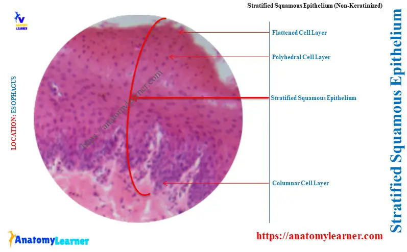
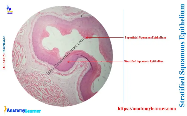
Lining epithelium of esophagus (nonkeratinized stratified squamous):
The nonkeratinized stratified squamous epithelium lines the lamina epithelium of the animal esophagus. The diagram shows the nonkeratinized stratified squamous epithelium from the esophagus (tunica mucosa layer).
The key features of the stratified squamous esophagus are –
- It shows multiple layers of cells, where the superficial cells are only flattened shaped,
- The cells of the basal layer of the esophagus show a columnar shape,
- The middle layer of the stratified squamous epithelium of esopahgus shows the polyhedral shape,
- The nuclei are oval in the basal layer, rounded in the middle layer, and transversely elongated in the superficial layer,
- There is no evidence of the keratin substance on the lining of esophagus,
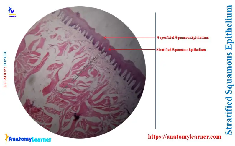
Lining epithelium of tongue (nonkeratinized stratified squamous):
The nonkeratinized stratified squamous epithelium covers both surfaces of the animal’s tongue. Here, the diagram shows the nonkeratinized stratified squamous epithelium from the animal tongue.
The dorsal surface of the tongue shows numerous projections or papillae with the stratified squamous epithelium. But, the ventral surface has no such projections or papillae.
It shows the multiple layers of cells – basal, middle, and superficial cells. The superficial cell layer of the tongue is flattened and shaped with the flattened nuclei.
You may know the details of the other structures from the tongue histology slide rather than the epithelium.
Keratinized stratified squamous epithelium location
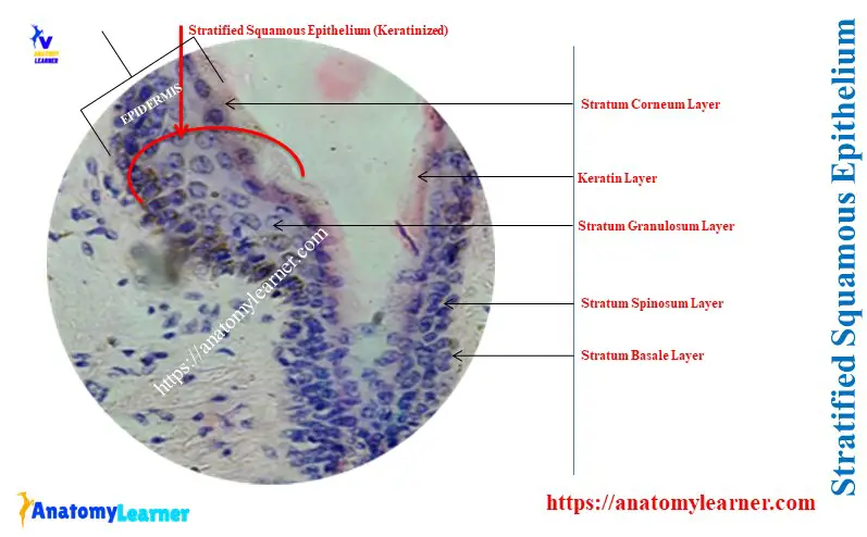
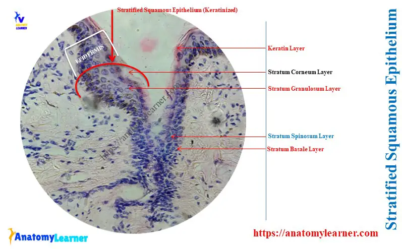
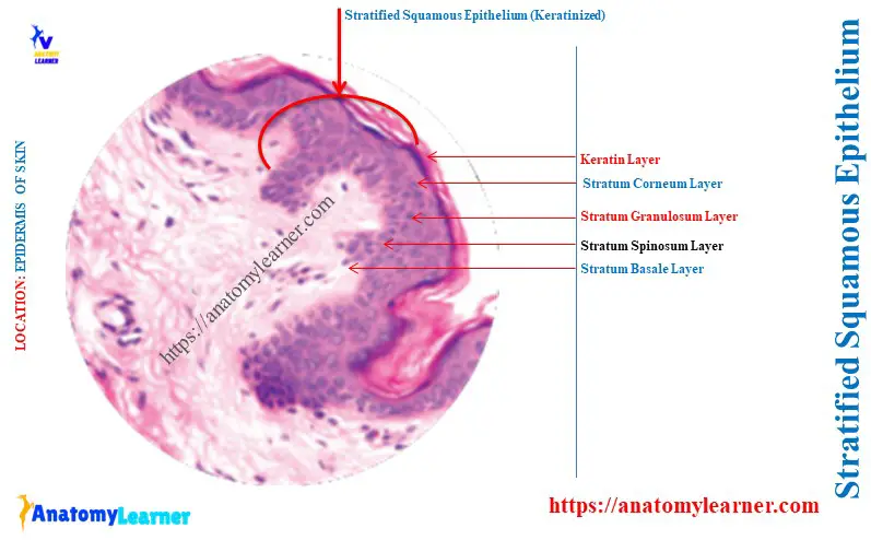
The typical features of the keratinized stratified squamous epithelium are found in the epidermis of the skin. The animal’s thick and thin skin represents the keratinized stratified squamous epithelium on their epidermis.
The diagram shows the keratinized stratified squamous epithelium from the animal’s thick skin. You may know more about the other features of skin from the skin histology slide.
There are distinct five cell layers in the stratified squamous epithelium of the skin –
- The deepest cell layer is the stratum basale: located next to the base membrane. This layer consists of a single layer of cuboidal or columnar cells.
- Stratum spinosum cell layer: consists of several layers of polyhedral cells. This cell layer is present only in the keratinized stratified squamous epithelium.
- Granular cells layer or stratum granulosum: consists of lamellar granules in their cytoplasm.
- Stratum lucidum cell layer: consists of eleidin and occurs only in the nonhairy skin of the animals.
- The outermost layer is the stratum corneum: consisting of keratinized, nonnucleated cells.
Unique features of the stratified squamous epithelium cell layers
The cells of the basal layer of the epidermis are mitotically active. They give rise to the cells that move towards the upper layer of the epithelium.
The cell surface of the spinosum layer possesses the small spiny processes. These appearances give rise to the name of this layer, stratum spinosum.
The spiny processes of the spinosum cells contain the tonofilaments. They remain condensed at the site of the desmosome (cell junction).
The cells of the stratum spinosum move towards the superficial surface and become more flattened. They accumulate keratohyaline and lamellar granules in their cytoplasm. This is the cells of the stratum granulosum of the epidermis of the thick skin.
The cells of the stratum lucidum contain a single layer of flattened cells between granulosum and corneum layers. These cells have a translucent appearance as they contain the eleidin.
The cells of the stratum corneum become the keratinized state by the keratinization process. This cell becomes nonnucleated and produces keratin substance above the surface.
Conclusion
So, you got the answer to the question – ‘Where is stratified squamous epithelium found?’. Two types of stratified squamous epithelium are found over the surfaces of the animal body subjected to friction.
The typical nonkeratinized stratified squamous epithelium may be found in the tongue, oesophagus, and laryngopharynx. Again, the keratinized stratified squamous epithelium is seen on the epidermis, nostril, and hard palate.