
Thyroid gland is a highly vascular bilobed endocrine gland that located at the lower part of neck inferior to the larynx. In thyroid gland histology you will find stroma (connective tissue framework) and parenchyma (thyroid follicles).
Hi dear anatomy learner, do you want to know about the thyroid gland histology? In this article I am going to share the basic thyroid gland histology with slide pictures and labeled diagram.
I will also share the thyroid gland slide drawing with you so that you might practice. If you need thyroid gland histology ppt then let me know.
After completing this article you will able to identify thyroid glands histology slide under microscope. I will enlist the important identifying characteristics of thyroid histology slide.
So, if you want to learn the basic of thyroid gland histology and identify it under microscope then you may continue this article.
I will also provide little information on thyroid gland anatomy. Okay, let’s get into the main part of today’s article – thyroid gland histology with labeled diagram.
Thyroid gland histology
From thyroid gland histology you might identify some important structures. First I would like to enlist these important structures that should be identifying under light microscope at laboratory. You might find out these structures from the thyroid slide pictures and labeled diagram.
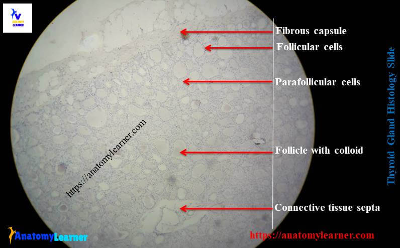
#1. Thin connective tissue capsule of thyroid gland
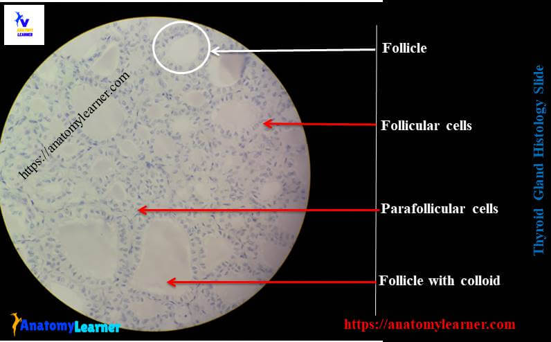
#2. Thyroid follicles with colloid
#3. Thyroid follicular cells of thyroid gland
#4. Connective tissue septa or trabeculae of thyroid gland or inter-follicular connective tissue
#5. Parafollicular cells of thyroid gland
#6. Arteriole and capillaries on connective tissue septa
Identifying characteristics of thyroid slide
Do you want to identify the thyroid gland histology slide under light microscope? Fine, hope these identifying characteristics of thyroid gland slide will help you a lot.
#1. Presence of numerous lobules that separated by dense connective tissue septa
#2. There are numerous and various sized follicles in each lobule
#3. Follicles are lined by simple cuboidal (varies on condition) epithelium (known as follicular cells)
#4. Thyroid follicles are filled with the colloid substances
#5. Presence of parafollicular or clear cells (C cell) in between the thyroid follicles. These cell occurs in single or clump on the periphery of the follicles.
#6. There are numerous blood vessels (arterioles, capillaries and venules) on the thin connective tissue septa
Hope these identifying characteristics will help you to identify the thyroid gland slide under light microscope.
Histology of thyroid gland with labeled diagram
Do you want to learn the details histology of thyroid gland with labeled diagram? Fine, I will describe capsule, stroma and parenchyma from the thyroid glands histology slide.
Stroma of thyroid histology
The stroma of thyroid gland histology includes capsule and interlobular thin and dense connective tissue. Thyroid gland is surrounded by a thin capsule of dense irregular connective tissue. This capsule extends into the parenchyma of thyroid gland and divides the gland into different lobules that contains thyroid follicles.
The intralobular loose areolar and reticular tissue provide the bed for the parenchyma where the thyroid follicles are present.
Follicle and follicular cell histology
In each lobule of thyroid gland you will find numerous and variable sized thyroid follicles that are filled with colloid substance. These follicles are lined by simple cuboidal epithelium (but vary with active or inactive condition of thyroid gland).
In resting condition the thyroid follicles are lined by simple epithelium (low cuboidal or even squamous epithelium) and colloid stained uniformly. But in active condition, these cells become cuboidal or columnar epithelium and colloid stained non-uniformly; these cells known as follicular cell. These follicular cells are responsible for synthesize and secret colloid and thyroid hormones.
You will also find the para-follicular or clear or C cell in between the thyroid follicles. These parafollicular cells are larger than the follicular cells and pale stained. Again these parafollicular cells occur as a single cell or in clump at the periphery of the follicles of thyroid gland structure. These cells have numerous small granule and responsible for secretion of calcitonin.
Do you want to know about the follicular cells of thyroid gland? Well, the cytoplasm of the follicular cells contains a round centered nucleus, rough endoplasmic reticulum at the basal part, numerous lysosome and secretory granules. You will also find the moderate number of microvilli at the apical pole of follicular cell membrane
Thyroid gland slide image
I am going to share some thyroid gland histology real slide so that you might learn and identify the important structures from thyroid structure.
If you need more thyroid slide images then you may follow anatomy learner at here to get update slide pictures.
Thyroid gland histology drawing
You might practice to thyroid gland histology drawing. Hope this drawing will help you to understand the structure perfectly. You might try to draw better thyroid slide image drawing then me.
Thyroid gland anatomy
If you want to know thyroid gland anatomy then you might read the article from the gross anatomy learning part. I am going to provide the basic anatomy of thyroid gland in animal. You know these are the bilobed (right and left) and highly vascular gland located at the lower part of the neck just inferior to larynx. The right and left thyroid gland are connected by median isthmus. They are brown red in color and heavier in female animal.
If you want you may learn other different endocrine gland histology with anatomy learner –
#1. Histological features of pituitary gland with real slide and labeled diagram
#2. Identification of adrenal gland histology structure – cortex and medulla of adrenal gland
#3. Histological characteristic of mammary gland of animal
Conclusion
Hope this guide is enough to understand the thyroid gland histology. If you want to know more about thyroid gland histology then let me inform.
I hope you will able to identify the histology of a thyroid glands slide under light microscope with the proper identifying characteristics.
If you think this is the best guide to identify thyroid histology slide under light microscope then share this article with your friends who want to learn thyroid gland histology with labeled diagram.
Thank you so much for staying with anatomy learner; don’t forget to follow anatomy learner at social media for more update.