The exact location of the dog’s liver is essential for veterinary practice. Here, you will know the answer to the question – ‘Where is the liver in a dog?’
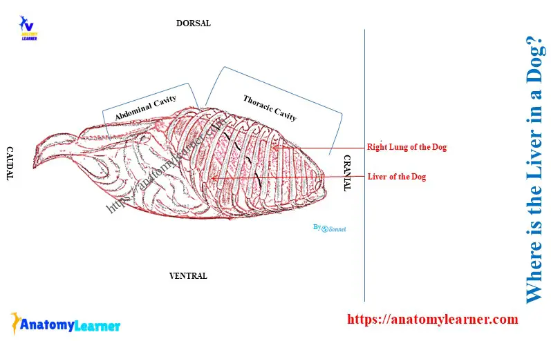
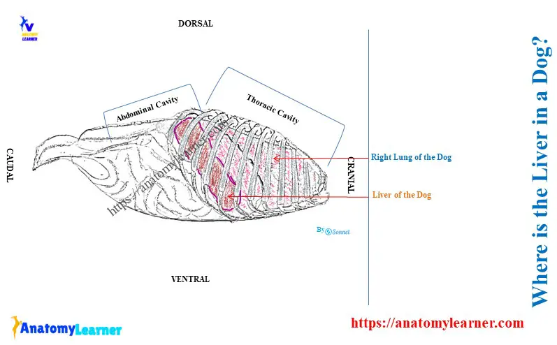
Quick answer: the dog’s liver is located at the right side of the abdominal cavity behind the diaphragm. It extends obliquely downward and forward from the last rib to the seventh rib on the right side of the body.
Here, I will show you the topographic location of the different lobes of the dog liver with diagrams. Thus, you will compare the location and features of other animals’ livers with the dogs’.
Where is the liver in a dog?
A dog’s liver is larger, usually weighing about three percent of its body weight. It is the dog’s largest gland and possesses both the exocrine and endocrine functions.
The dog’s liver is situated on the right side of the abdomen behind the diagram. It also has a relationship with the stomach, duodenum, pancreas, and right kidney.
Here, the diagram shows the location of the dog’s liver on the right side of the abdomen with other organs.
To understand the topographic position of the dog liver, you might know its anatomy details. You might have an idea of the dog liver’s surfaces, borders, lobes, and processes.
Overview of the dog liver
So, first let’s see how many surfaces, borders, lobes, and processes a dog’s liver has. Here, Table 1 shows the lobes and processes of the dog liver –
| Dog liver Lobes | Name of the Sublobes | Name of the Processes |
| Left hepatic lobe | Left lateral hepatic lobe Left medial hepatic lobe | |
| Quadrate lobe | ||
| Right hepatic lobe | Right lateral hepatic lobe Right medial hepatic lobe | |
| Caudate lobe | Papillary process Caudate process |
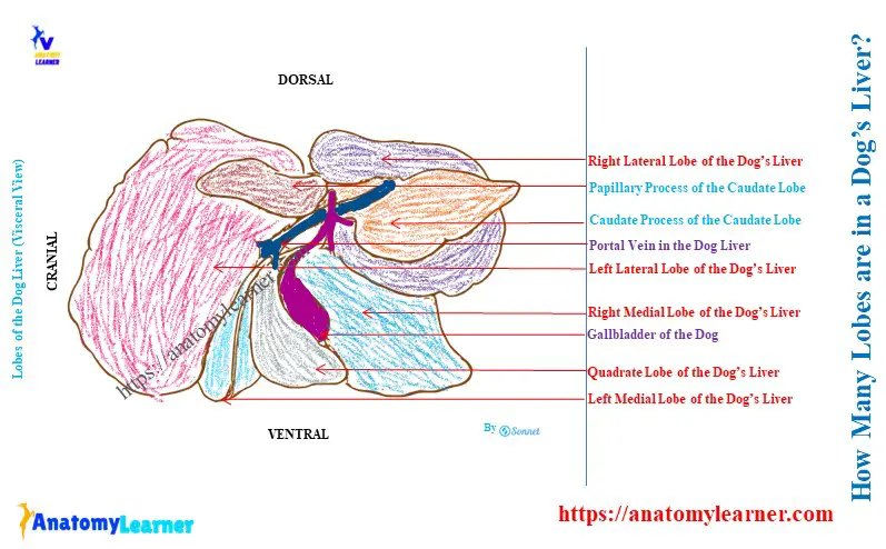
Thus, the dog’s liver has 4 lobes, 4 sublobes, and 2 processes. Here, the diagram shows the dog liver anatomy’s different lobes, sub-lobes, processes, and surfaces.
The dog liver has two surfaces – strongly convex diaphragmatic (parietal) and irregular concave visceral surfaces. Again, this liver shows the dorsal, ventral, right, and left borders on its anatomy.
You will also see some impressions and porta on the structure of a dog liver. The major impressions on the dog liver are the gastric, duodenal, and renal. But, you will find more impressions on the structure of a cow liver.
Now, I will describe the relationship of a dog liver’s surfaces, borders, and lobes with other different organs. Thus, you will easily understand the relative position of the dog’s liver.
Position and relation of the surfaces of dog liver
The diaphragmatic surface of the dog liver is strongly convex in all directions. This surface lies in contact with the diaphragm and possesses three distinct parts –
- Right part: it is the largest and extends at the level of the sixth intercostal space to the last intercostal space,
- Cranial part: it is located at the left side of the median plane and contains a shallow, broad, and indistinct cardiac impression,
- Dorsal part: This part is located at the median plane and is deeply notched by the caudal vena cava and esophagus. Here, the caudal vena cava lies to the right of the esophagus on the dorsal groove of the diaphragmatic surface.
The visceral surface of the dog liver is in contact with the stomach, duodenum, pancreas, and right kidney. You will also see the gallbladder at the irregular concave visceral surface of the dog liver.
But, the most prominent feature on the visceral surface of the dog liver is the papillary process. And you know, the papillary process is the medial extension of the caudate lobe of the liver.
All the organs connected with the visceral surface of the dog liver produce little impression. Typically, the gastric impression (made by the stomach) occupies the whole left half of the visceral surface. However, this impression is variable due to the different conditions of the dog’s stomach.
The duodenal impression is located between the junction of the right and quadrate lobes on the visceral surface. The right kidney on the caudate process of the caudate lobe forms a deep and hemispherical fosse.
Location and relation of the lobes of a dog liver
The left hepatic lobe of the canine liver lies almost to the left of the median plane. Here, the left lateral part of the left hepatic lobe begins dorsally deep to the left crus of the diaphragm.
Again, the visceral surface of the left lateral hepatic lobe lies on the fundus of the dog’s stomach. The central part of this sub lobe lies adjacent to the lesser omentum of the dog.
The left medial hepatic lobe is roughly triangular and separated from other lobes by fissures. Here, the left medial and lateral lobes are joined by a deep, narrow bridge of liver tissue.
The quadrate lobe of the dog liver lies primarily in the median plane. Sometimes, the middle of the visceral surface of the quadrate lobe contributes to forming the fossa for the gallbladder.
A dog liver’s small right hepatic lobe lies totally to the right of the median plane of its body. Here, the medial part of the right hepatic lobe fuses with the medially locates quadrate lobe. The medial surface of the medial right hepatic lobe contributes to forming the right half of the fossa for the gallbladder.
The right lateral hepatic lobe overlaps with the caudate process of the caudate lobe. It also relates to the lateral to the caudal vena cava.
The lesser omentum encloses the pyramidal papillary process of the caudate lobe. This process lies in the lesser curvature of the dog’s stomach.
The caudate process of the caudate lobe forms the most caudal part of the dog liver. This process lies on the right side of the median plane and extends up to the last intercostal space.
How is the dog’s liver fixed in its position?
The dog’s liver is entirely covered by the peritoneum except where the gallbladder is attached. This layer of the liver forms the serous coat.
Again, this serous coat fuses with the underlying subserous layer and fibrous capsule. The fibrous capsule of the dog liver is the thin but dense layer of collagen tissue.
The continuation of the serous and fibrous coat forms the coronary and several folds. They provide parietal attachment on the dog’s liver.
You will find a 5 ligaments that help the dog’s liver fix in its position. These five ligaments of the dog liver are –
- A coronary ligament of the dog’s liver,
- The right triangular ligament of the dog liver,
- A left triangular ligament of the dog liver,
- The falciform ligament of the dog liver and
- A hepatorenal ligament of the dog liver,
The coronary ligament is triangular in outline and reflects the close embryonic relationship between the diaphragm and liver. Here, the right triangular ligament extends between the diaphragm and the dorsal part of the right lateral lobe.
The left triangular ligament of the dog liver has several components. Here, the cranial component of the left triangular ligament runs from the left lateral lobe to the diaphragm.
The caudal component is more significant and contain the fibrous appendix of the liver. Here, the fibrous appendix is the thin and narrow part near the free border of the liver.
Finally, the falciform ligament of the dog liver extends between the liver and diaphragm. It also extends from the ventral body wall to the umbilicus.
The hepatorenal ligament extends from the medial part of the renal fossa to the ventral surface of the right kidney.
Conclusion
So, the liver is located on the right side of the dog’s abdominal cavity. The diaphragm is located cranial to the dog’s liver.
The dog liver’s caudal part and visceral surface relate to the stomach, pancreas, duodenum, and right kidney. Five different ligaments and a serous coat help the dog’s liver to remain in its position.