The femur of a cat is a long bone of the hind limb. Its osteological features are a little different than those of the dog’s femur bone. Here, you will learn the details of the osteological features of the cat femur bone with the diagram.
Quick answer: The cat femur is the hindlimb’s long bone that possesses a cylindrical shaft and two extremities. Here, the proximal end contains the head, neck, trochanters, and fossa, whereas the distal end contains condyles, trochlea, and fossa.
I will identify and describe all the osteological features from the shaft and extremities of the cat femur bone.
What is a femur on a cat?
The femur is the largest and most massive bone of the cat’s skeleton. It forms the leg segment of the cat’s hindlimb.
The direction of the cat femur is similar to the direction of the ox femur bone. It also directed obliquely downward and forward.
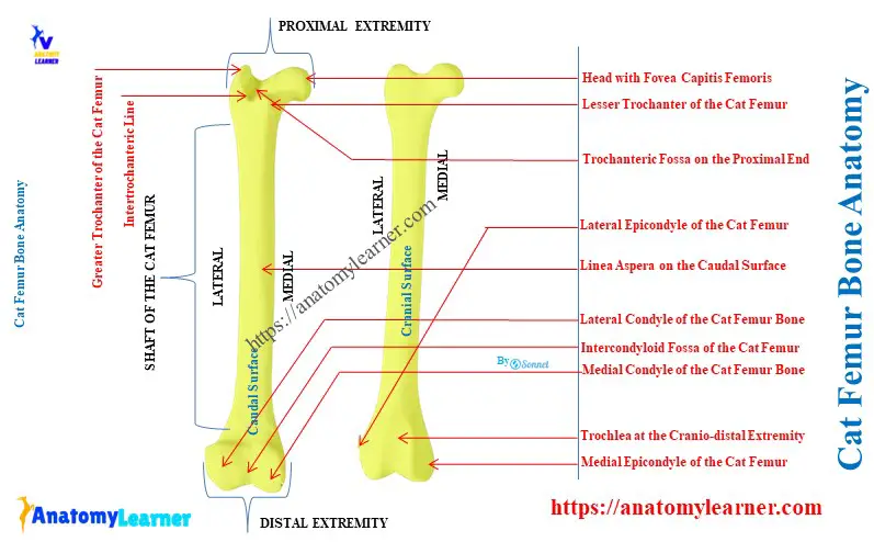
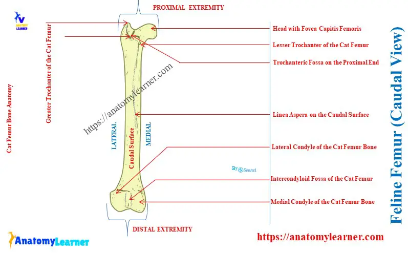
Here, the diagram shows the femur bone from the hind limb of a cat. The shaft and two extremities (proximal and distal) are identified in the cat’s femur bone labeled diagram.
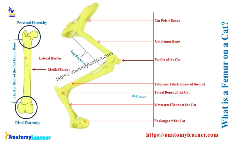
This cat’s femur bone is located between the hip and tibia bones. The proximal part of this cat’s femur joins with the pelvic bone with the hip joint. Again, distally, it joins with the tibia bone by the stifle joint.
The inclination of the feline femur bone on the horizontal plane is about 70 – 80 degrees. As a typical long bone, this femur also represents –
- A cylindrical shaft or body (that contains various surfaces and borders) and
- The proximal and distal extremities of the feline femur bone,
Cat femur bone anatomy
In the cat femur bone anatomy, you might describe the features of the shaft and extremities individually. Here, the shaft of the femur is an elongated or cylindrical structure that possesses 4 surfaces and 4 borders.
But, the surfaces of the femur bone continue with each other. Thus, the area of the 4 surfaces is hard to identify specifically.
Again, four borders are practically absent from the shaft of the cat’s femur bone. You will clearly identify only two borders from the cat’s femur (lateral and medial borders).
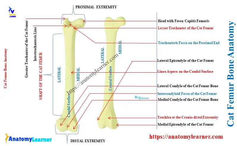
The diagram shows all the surfaces and borders of the feline femur bone. It also shows the features from the proximal and distal extremities of the feline femur bone.
The proximal extremity of the cat’s femur contains the following osteological features –
- Medially located head with fovea capitis femoris,
- Cranio-medially located neck of the femur,
- Laterally located greater trochanter,
- Caudo-medially located lesser trochanter,
- Intertrochanteric crest or line (continuation of the greater trochanter),
- Proximally located trochanteric fossa (in between the greater and lesser trochanters),
The distal extremity of the cat’s femur bone contains the following osteological features –
- More prominent lateral and smaller medial condyles,
- Caudally located intercondyloid fossa (between lateral and medial condyles),
- Prominent medial epicondyle and smaller lateral epicondyle,
- Cranially located trochlea for the patellar articular surface,
Summarize the osteological features of cat femur bone
Table 1 shows the summary of the osteological features from the shaft and extremities of the cat femur –
| Cat Femur Bone Anatomy | Osteological Features |
| Shaft of the femur | Four surfaces – Cranial surface Caudal surface Medial surface, and Lateral surface Two borders – Lateral border, and Medial border, |
| Proximal extremity of the femur | Ball-like head Irregular fovea capitis femoris Constricted neck Prominent greater trochanter Smaller lesser trochanter Distinct intertrochanteric line or crest Moderate trochanteric fossa |
| Distal extremity of the femur | Prominent lateral condyle Prominent medial condyle Irregular lateral epicondyle Irregular medial epicondyle Caudal intercondyloid fossa Cranial trochlea |
Shaft of a cat femur bone
The cranial, medial, and lateral surfaces of the shaft of the feline femur bone are continuous. But, the caudal surface of the shaft is a little wide and smooth.
You will see a linea aspera (ridge) on the caudo-medial surface of the femur’s shaft. It is a less conspicuous line that extends from the lesser trochanter of the proximal end. This ridge provides the site for strong muscle attachment.
The medial border of the shaft possesses a smaller trochanter than the greater trochanter. This is the lesser trochanter of the cat’s femur bone.
But, the lateral border of the shaft doesn’t possess any distinct supracondyloid fossa and tuberosity. The shaft of the dog femur bone also doesn’t contain any supracondyloid fossa and tuberosity on the lateral border.
You will find the developed supracondyloid fossa and tuberosity on the lateral border of the horse and ox femur bones.
So, the key features of the shaft of the feline femur bones –
- Presence of a linea aspera or ridge on its caudomedial surface, and
- Absence of the supracondyloid fossa and tuberosity on the lateral border,
Proximal extremity of the feline femur bone
Head and neck of the femur:
There is a prominent ball-like projection of the femur on its proximal extremity. This is the head of the cat’s femur bone that faces medially and makes the hip joint with the acetabulum of pelvic bones.
Here, the head of the femur joins with the articular surface of the acetabulum. Again, the fovea capitis femoris is the site of attachment of a round ligament. This ligament helps to bind the femur bone with the ventral part of the acetabulum (within the acetabular fossa).
At the medial aspect of the head, you will see an irregular depression. This irregular depression on the surface of the head is the fovea capitis femoris. But, it doesn’t show any distinct notch on the margin of the fovea capitis femoris structure like the horse femur.
The cranio-medially directed constricted part of the head is the neck. It articulates the head of the feline femur bone with the epiphysis bone.
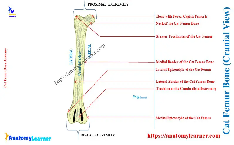
Trochanter on the proximal extremity:
The larger bony projection on the lateral aspect of the proximal extremity of the femur is the greater trochanter. Within this greater trochanter, the muscles of the cat’s hip are attached.
The greater trochanter of the cat’s femur bone remains at the level of the head. But, it extends above the level of the head in the case of the ox and horse femur bones.
At the medial aspect of the greater trochanter, a conspicuous depression is found. This is the trochanteric fossa of the feline femur bone.
The caudomedial aspect of the proximal extremity shows a comparatively small trochanter. In between these two trochanters of the femur, you will see a crest or line. This is the trochanteric crest (or sometimes line) of the cat’s femur bone.
Several ridgelike structures are running around the constricted neck and lesser trochanter. The intertrochanteric ridge is the prominent ridge on the caudal aspect of the proximal extremity.
This intertrochanteric ridge also continues the caudomedial aspect of the femur bone from the lesser trochanter. Then, this ridge continues with the linea aspera of the shaft of the feline femur bone.
The distal extremity of the feline femur bone
Cranial aspect of the distal extremity:
The presence of the trochlea is the most prominent osteological feature at the cranial aspect of the distal extremity. It is tongue-shaped and has a smooth surface for articulation with the caudal surface of the patella.
Here, the patella is a small triangular sesamoid type of bone in the stifle joint of a cat. This patella in a cat is comparatively more significant than the other animals. Again, the cranial surface of the cat’s patella is more convex than that of other animals’ patellas.
There are some grooves (lateral and medial) present on the cranial surface of the distal extremity. The ligaments of the stifle joint and some of the tendons pass over these grooves.
Again, there is some nutrient foramina in the distal extremity of the cat femur. Different vessels and nerves pass through these foramina of the cat’s femur bone.
Caudal aspect of the distal extremity:
The most prominent feature at the caudal-distal extremity of the feline femur bone is the condyle. There are prominent lateral and medial condyles on the corresponding aspect of the femur at its distal extremity.
Both two condyles have smooth and rounded articular surfaces. They articulate with the proximal articular surface of the cat’s tibia bone.
In between these two condyles, there is a deep posterior notch. This is the intercondyloid fossa that separates the condyles.
You will also see irregular shape bony prominence at the corresponding site of two condyles. These are the epicondyles, termed the lateral and medial epicondyles, of the cat’s femur bone.
Both epicondyles are located just above the corresponding condyles. They both provide the site for the attachment of the tendon of the muscles.
Key features of the extremities of a cat femur
- The proximal extremity has 5 structures: head, neck, trochanter, trochanteric fossa, and trochanteric crest, and
- The distal extremity has 5 essential features: Condyles, epicondyles, trochlea, and intercondyloid fossa,
How do you differentiate a cat’s femur from an ox’s and horse’s femur bones?
Answer: you may easily differentiate the cat femur bone from the ox and horse femur by its size and external features. The body of the ox and horse femur bone possesses supracondyloid fossa and tuberosity on its distal extremity. But, the feline femur bone doesn’t possess any supracondyloid fossa and tuberosity on its body.
Again, the horse femur bone possesses an exceptional third trochanter on the lateral aspect of the body. The fovea capitis femoris of a horse femur bone is notched, whereas the cat’s fovea is very small and free of any notches.
The trochanteric ridge or line of the feline femur is directed obliquely. But, in the horse femur, it is directed vertically along the length of the bone.
In both the ox and horse femur, the head is located below the level of the greater trochanter. Again, the greater trochanter of both ox and horse femur bone divides into cranial and caudal parts.
Conclusion
So, the cat femur bone forms the leg segment of the hindlimb and contains all the typical features of a long bone. The shaft of the femur is smooth, cylindrical, and possesses linea aspera.
Both the proximal and distal extremities of a cat femur possess unique osteological features. The proximal head, trochanter, distal trochlea, and condyle are clinically essential structures of the femur bone.