The hindlimb of a dog divides into four different regions, like the forelimb. Here, you will get the answer to the question – ‘ what are the regions of the hindlimb of a dog?’
Quick answer and explanation: the pelvic girdle, thigh, leg, and pes are the four regions or segments of the dog’s hindlimb. The bones of the hindlimb of a dog form these four specific regions.
Here, I will show you the four different regions of the dog’s hindlimb with their components. Thus, you will practically identify these regions from the live dog after completing this article.
What are the regions of the hindlimb of a dog?
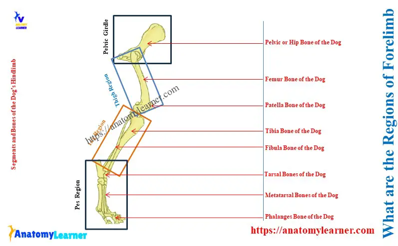
Explanation of four segments of a dog’s hindlimb: the hindlimb of a dog consists of pelvic bones, femur, tibia, fibula, carpals, metacarpals, and phalanges. You will find the following four regions or segments of the hindlimb of a dog –
- Pelvic girdle (bony pelvis),
- Thigh region,
- Leg region, and
- Pes region,
Here, the diagram shows these four regions with the bones from the dog’s hindlimb. You will find more diagrams on the specific regions of the dog’s hindlimb in the next section of this article.
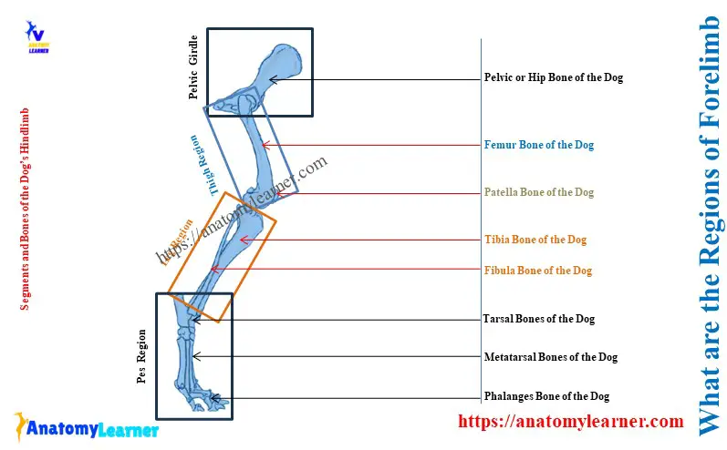
Table 1 shows the bone’s involvement in the four different regions of the hindlimb of a dog –
| What are the regions of the hindlimb of a dog | Bones are involved in the regions of the hindlimb |
| Pelvic girdle | It consists of the pelvic bones, sacrum, and first one or two caudal vertebrae. |
| Thigh region | The thigh of a dog consists of the femur bone. |
| Leg region | Dog’s leg includes the tibia and fibula bones. |
| Pes region | It consists of tarsals, metatarsals, and phalanges |
Pelvic girdle segment of a dog
The right and left os coxae of the dog’s pelvic bone form the ossa coxarum. Here, each os coxae of the dog consists of the ilium, ischium, and pubis bones.
The ossa coxarum with the sacrum and first one or two caudal vertebrae form the pelvic girdle. Thus, three bones are involved in forming the dog’s pelvic girdle – hip bone (pelvic bone), sacrum, and caudal vertebrae (first two).
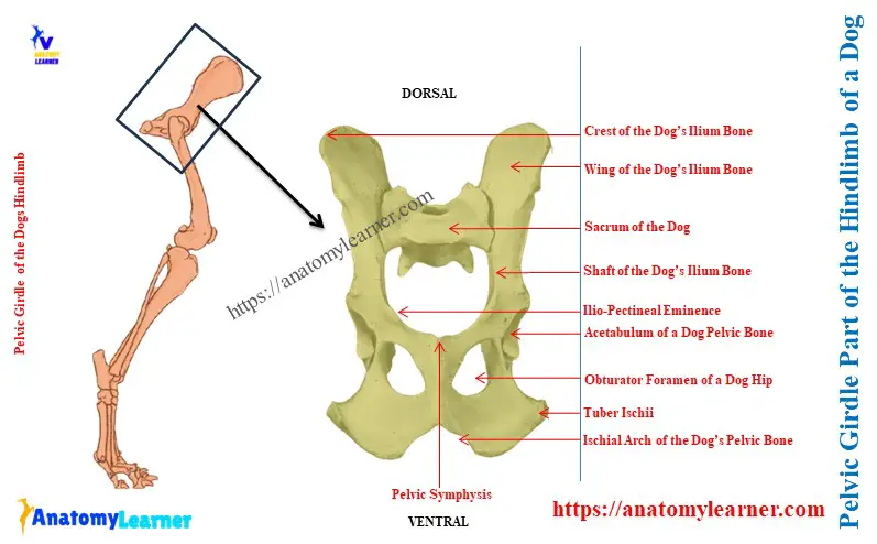
So, the pelvic girdle of a dog = hip bone + Sacrum + first one or two coccygeal vertebrae.
Features of the dog sacrum and caudal vertebral bones
The anatomy of the dog sacrum is structurally different than that of the ox sacrum. In the dog, there are only three sacral vertebrae fuses from the sacrum bone.
The dog sacrum possess two dorsal and ventral sacral foramina. It shows a wide articular surface on the wings. The longitudinal median sacral crest of the dog shows a notch in its middle.
The first and second caudal vertebrae of the dog’s vertebral column are more well-developed than those of the ox. You will find a great variation in the number of the caudal vertebrae that form the dog’s tail.
Dog’s pelvic bones (hip bones)
Here, the dog’s pelvic bones greatly vary from the ox hip bones. You will find a detailed description of the dog’s pelvic bone here on Anatomylearner. Thus, I want to provide the identifying features of the dog’s pelvic bones now.
Here, the iliums of the dog’s pelvis are parallel to each other. The crest of the ilium is convex, and the gluteal surface is strongly concave.
The ischium bone of the dog’s pelvis is twisted. You will find the great difference in the features of the ischial tuberosity of the dog’s ischium bone. Here, the ischium tuberosity of the dog ischium is flat.
The ischiatic spine is blunt in the dog’s hip. Again, you will see a shallow depression on the greater and lesser ischiatic notches.
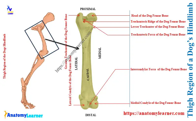
The thigh region of a dog
The femur (round bone) forms the thigh region of a dog’s hindlimb. There are fewer identifying features for the dog’s femur bone compared to the ox femur.
Here, the greater trochanter of the dog’s femur bone is at the bottom level of the head. The cranial and caudal segments of the greater trochanter are hardly identified in dogs. Again, the lesser trochanter of the dog’s femur bone is in the form of a tubercle.
The trochanteric ridge is oblique, and the fossa is less deep compared to the ox. There is no supracondylar fossa at the distal extremity of the body of the dog’s femur.
The leg region of a dog
The tibia and fibula of a dog from the leg region. Here, the body of the dog’s tibia is prismatic at its upper part. Again, the body of the dog’s tibia is cylindrical at its lower part.
You will find the facets on the lateral aspect of the cylindrical part of the tibial body for articulation with fibula bone. Here, the tibial crest is very prominent in the dog’s tibia bone.
The fibula bone of a dog is comparatively thin and extends through the whole length of the tibia bone. Here, the distal end of the dog’s fibula bone is thick.
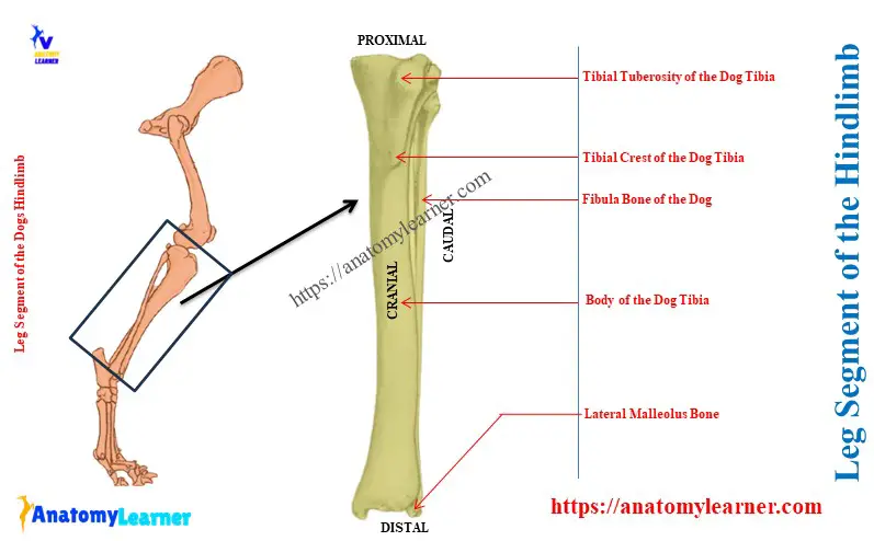
Suggested article from anatomy learner: dog tibia anatomy – canine leg bone, muscle, and vessels with the diagram.
Pes segment or region of a dog
Seven tarsals, five metatarsals, and fourteen phalanges form the pes segment or region of a dog’s hindlimb.
The seven tarsal bones of a dog are arranged into three rows –
- First row consists of the tibial tarsal (medially) and fibular tarsal (laterally),
- Second row consists of the central tarsal bone, and
- Third row consists of the first, second, third, and fourth tarsal bones (from medial to lateral),
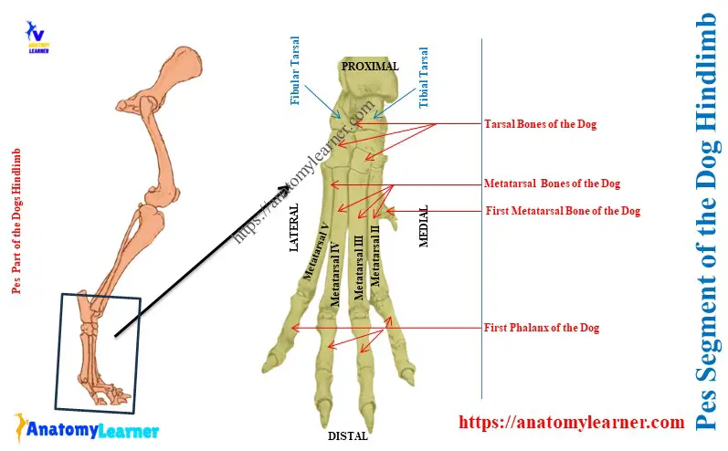
Except for the first metatarsal, all other metatarsals are well-developed. Here, the first metatarsal of the dog’s pes is in the form of a cone. You will find a detailed guide to the dog tarsal and metatarsal bones in another article by Anatomylearner.
From second to fifth digits of a dog consist of three developed phalanges (proximal, middle, and distal). But, the first digit of a dog consists of two phalanges.
How many bones does a dog have in its hindlimb?
Answer: There are a total of five bones in each hindlimb of a dog. But, the main bones of the dog’s hindlimb are thirty. The other fifteen are the sesamoid bones of the hindlimb.
Again, the hip or pelvic bone of a dog consists of ilium, ischium, and pubis. Thus, there are a total of three bones in both the right and left pelvis. As all these bones remain fused, they are considered as a single bone.
Table 2 shows the total number of various bones from the dog’s hindlimb –
| Bones of the hindlimb of a dog | Number of the bones in a dog’s hindlimb |
| Pelvic bone | 1 |
| Femur bone | 1 |
| Patella (sesamoid bone) | 1 |
| Tibia and Fibula bones | 2 |
| Tarsal bones | 7 |
| Metatarsal bones | 5 |
| Phalanges in hindlimb | (4 x 3) + 2 = 14 |
| Sesamoid bones in a dog hindlimb Proximal sesamoid bones Distal sesamoid bones | (4 x 2) + 1 = 9 (5 x 1) = 5 |
| Total bones of the forelimb | 45 |
The sesamoid bones of the dog’s hindlimb are fifteen, which is more than the forelimb. This is because there is a large sesamoid bone (patella) in the stifle joint of a dog’s leg.
Conclusion
So, the pelvic girdle, thigh, leg, and pes are four regions of the hindlimb of a dog. These four regions are also termed as the segments or parts of the hindlimb. Now, you might practically identify these four regions from the hindlimb of a live dog.