
Lymph node is a oval or bean shaped structure located along the extensive drainage system of lymph vessels of animal. In lymph node histology, you will also find two main components – the connective tissue thin framework and parenchyma.
Hi there, welcome back to anatomy learner and thank you so much for getting into this article. If you want to learn the details histology of lymph node from animal then this article is for you. Okay, in this article, I am going to describe the histological features of lymph node along with their identification characteristics.
I will also show you the every single structures from the cortex and medulla of lymph node histology real slide images. You will get full histological features of cortex and medulla of lymph node with labeled diagram. At the end, you will also get lymph node histology drawing images.
Fine, want to continue this article and learn the histological features of lymph node structure? Let’s start tp learn the histological features of lymph node cortex, paracortex and medulla.
Lymph node histology
You know, lymph nodes are the only lymphatic organs that contain both afferent and efferent lymphatic vessels. In lymph node, you will find the hilus – where blood or lymph vessels enter or leave the lymph node. There are cortex (contains lymphatic nodules, diffuse lymphatic tissue), paracortex (contains lymphatic tissue) and medulla (contain lymphatic tissue and arranged in cords) in the parenchyma of lymph node histology slide.
Before going to the details histological features of lymph node I would like to enlist the most important structure that you might identify from lymph node slide under the light microscope.
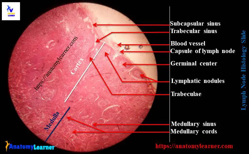
#1. Thin capsule of lymph node
#2. Thin trabeculae of lymph node
#3. Subcapsular sinus of lymphnode
#4. Trabecular sinus in lymph node
#5. Lymphatic nodules with germinal center in the cortex of lymph node histology
#6. Medullary cord in the medulla of lymph node structure
#7. Medullary sinus in the medulla of lymph node
Okay, let’s find out these important histological features from lymph node slide.
Identification points of lymph node slide
Do you want to identify the lymph node slide under light microscope? Well, hope the following important identifying characteristics will help you to identify lymph node slide under light microscope.
#1. Presence of thin dense irregular connective tissue capsule at the tissue section
#2. There are thin trabeculae extends from the thin capsule into the parenchyma of the tissue section as an irregular septa throughtout the organs
#3. The capsular sinus and trabecular sinus are present in the tissue section
#4. Presence of outer dark staining cortex and inner light staining medulla in the tissue section
#5. There are lymphatic nodules along with germinal center at the cortex of the sample section
#6. Presence of medullary cords and medullary sinus in the meduall region of this tissue section
So, this is a slide of lymph node.
Lymph node histology description with slide and labeled diagram
Fine, do you want to learn the details lymph node histology with real slide pictures and also with labeled diagram? Great, I am going to describe the every single parts of lymph node structure.
In thin connective tissue framework of lymphnode, you will find the following structures –
#1. Thin capsule of lymph node
#2. Trabeculae of lymphnode
#3. Reticular stroma of lymph node
Again, in the parenchyma of lymphonde you will find the following parts –
#1. Cortex of lymph node
#2. Para-cortex of lymph node structure
#3. Medulla of lymph node
Connective tissue framework of lymphnode
Lymph nodes are surrounded by a capsule primarily composed with dense irregular connective tissue. You will find few smooth muscle cells in the connective tissue capsule in ruminant lymph node.
Beneth the capsule there is subcapsular sinus that receive the afferent vessels and continue with the trabecular sinus. Again the trabecular sinus continue with the medullary sinus in lymph node structure.
The thin capsule sends numerous trabeculaes into the interior part of the lymphnode (both in cortex and medulla). The trabeculae provides support the entire lymph node and carries the blood vessels and nerves.
Again, the stroma of lymph node comprises of reticular cells and reticular fibers. You will also find the lymphocytes, macrophages and plasma cells in the reticular network of stroma of lymph node.
Cortex of lymph node structure
In the cortex of lymph node there are primary and secondary lymphatic nodules that seperated by diffuse lymphatic tissue. You know these lymphatic nodules are formed by the B lymphocytes (mainly) in lymph node. If you find a lymphatic nodues without a pale staining germinal center (contains larger lymphoblast) then the nodule termed as primary lymphatic nodule, whereas nodule with germinal center termed as secondary lymphatic nodule.
Para cortex is the inner cortical zone of lymph node and has no specific boundary. This paracortex mainly consists of T lymphocytes and termed as thymus dependent zone. The dense lymphoid tissue of para cortex becomes continue with the medullary cord of medulla of lymph node.
Medulla of lymph node histology
The medulla of lymph node histology is less organized than the cortex of lymph node. In the medulla of lymph node, you will find two important components – medullary cords and medullary sinus.
Medullary cords are the branching and anastomosing cords of typical lymphoid tissue; made with mainly T lymphocytes. You will also find the prominent plasma cells and also macrophage in the medullary cords in medulla of lymph node.
The medullary sinuses are atypical lymphoid tissue located between the medullary cords of lymph node structure.
Lymph node histology drawing
Now, I am going to share the lymph node histology drawing with you so that you might understand the every single structures from the lymph node. Hope you will also practice the drawing of lymph node slide images.
Do you need more images related to lymph node slide? Fine, you may follow anatomy learner at social media for more updated pictures of lymph node slide.
You might also like the following different organ’s histology from anatomy learner –
#1. Histological features of spleen – red and white pulp histology with real slide pictures
#2. Identification points of thymus under light microscope
Conclusion
I know you got the basic lymph node histology with real slide pictures and labeled diagram. Hope you will able to identify the lymph node histology slide under light microscope with the help of identification points that I provided.
If you want you may share this article with your friend who wants to learn lymph node histology with labeled diagram and also with the real slide pictures.
Don’t forget to follow anatomy learner blog to get more updated articles on veterinary anatomy and histology.