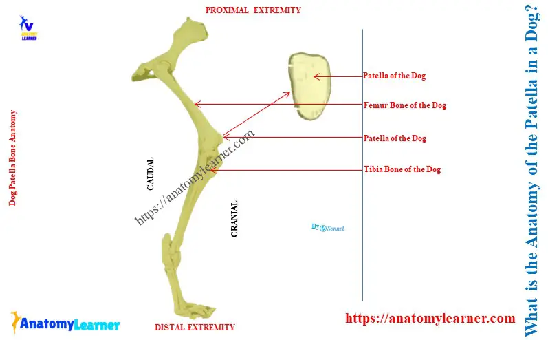
The dog patella is a triangular sesamoid bone placed in front of the trochlea of the femur. Here, you will know the answer to the question – ‘What is the anatomy of the patella in a dog?’
Quick answer: the anatomy of a dog patella consists of a base, an apex, two surfaces, three borders, and three angles. It is comparatively more extended than the ox or horse, and the cranial surface is more convex.
I will describe the position and details of the anatomical features of the dog patella with the diagram. Again, you will find a comparison among the patella of a horse, ox, and dog at the end of this article.
Key features of the dog patella anatomy
First, let’s see the diagram where I identified all the anatomical features of the dog patella. It mainly shows the following features of the dog patella –
- The proximal blunt base of the patella,
- Distal pointed apex of the patella,
- Cranial quadrilateral free surface,
- Caudal quadrilateral articular surface, and
- Lateral and medial borders of the patella,
Compared to other animal’s patella bones, you will find the below-mentioned key features in the dog patella –
- The dog’s patella is comparatively longer than other animal’s patella,
- A quadrilateral cranial surface of the canine patella is free and more convex,
- The caudal quadrilateral surface of the canine patella is concave along the length and convex across the width,
- A lateral and a medial angle are distinct from those of the horse patella,
Now, you will know the details of the anatomical features of the canine patella anatomy with diagrams.
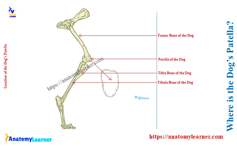
Where is the dog’s patella?
The dog’s patella is the most prominent sesamoid bone of the body. It is ovate, slightly curved, and located on the articular surface of the trochlea of the femur bone.
Actually, the caudal articular surface of the dog’s patella is concave. Thus, this concave surface articulates with the trochlea of the dog’s femur.
And you know, the femur is the hindlimb bone of the dog that forms the thigh segment. The patella of the dog’s hindlimb slides upward during the extension of the hindlimb. At the same time, it goes downward of the trochlear surface during the flexion of the hindlimb.
Here, the diagram shows the normal position of the dog’s patella on the trochlea of the femur bone.
What type of bone is the patella?
A patella is a sesamoid type of bone in the dog skeleton. There are different types of sesamoid bones in the dog skeleton –
Proximal sesamoid bone: includes the sesamoid bones that are located on the caudo-distal part of the metacarpal and metatarsal bones and
Distal sesamoid bones: these are sesamoid bones that are located between the middle and distal phalanges of both limbs.
Here, you will find two patellae on the knee joint of the dog’s body (one for each joint). They are the largest sesamoid and possess the typical features of the sesamoid bones.
You may know the total number of the dog’s sesamoid bones from the below-mentioned article –
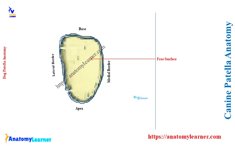
What is a normal patella in a dog?
A normal patella is a triangular or ovate-shaped structure on a dog. It articulates with the trochlea of the femur and presents two surfaces, three borders, three angles, and a base.
Here, the ventral angle of the dog’s patella forms the apex. It is a pointed structure of the patella bone. This apex of the dog’s patella faces distally.
Let’s see the overview of a normal patella on a dog –
- Shape: triangular or ovate shape, little curved bone,
- Features: two surfaces, three borders, three angles, and a base,
- Cranial surface: free, convex, and quadrilateral,
- Caudal surface: quadrilateral and articular surface for trochlea,
- Borders: both forms base dorsally and apex ventrally,
- Base: faces proximally and caudally, and
- Apex: pointed structure and faces distally,
What is the anatomy of the patella in a dog?
To describe the anatomy of the patella in a dog, you might cover the following –
- Features of the surfaces (cranial and caudal),
- Features of the borders (lateral and medial),
- Formation of the base and apex, and
- Attachment of the patella with the patellar ligaments and others,
Surfaces of the dog’s patella
Both the free and articular surfaces of the canine patella are quadrilateral. Here, the cranial free surface is rough for the muscular and ligamentous attachment.
Actually, this patella is an ossification in the tendon of insertion of the large extensor quadriceps femoris muscle. The name of this tendon is the patellar straight ligament. It attaches to the tibial tuberosity of the dog’s tibia bone to the specific part of the patella.
This patella alters the direction of the tendon of the quadriceps femoris muscle. It also provides a more excellent bearing surface for the tendon to play on the trochlea of the femur bone.
The caudal articular surface of the canine patella is smooth and convex in all directions. But, in some of the patella, it shows the longitudinal striation.
There are two or three parapatellar fibrocartilages present on either side of the caudal surface of the patella. These are the grooved cartilages that articulate with the ridges of the trochlea of the femur.
They also increase the articular surface of the trochlea of the femur bone. You may also see two other cartilages on the proximal extremity of the canine patella structure.
Borders, base, and apex of the canine patella
Both the lateral and medial borders converge ventrally to form the apex of the patella. It is slightly more pointed than the base of the patella. It does not extend beyond the articular surface of the canine patella.
Here, the medial angle of the canine patella is large, and the lateral angle is blunt. The medial border of the patella possesses several nutrient foramina.
The basis patellae (base) is blunt and faces proximally. It is convex transversely and concave craniocaudally.
How do you differentiate dog patella from ox and horse patella?
You will find major differentiating features in the shape, surface, base, and apex of the patella among ox, horses, and dogs. Here, table 1 shows the significant differences among the ox, horse, and dog’s patella –
| Features | Ox patella | Horse patella | Canine patella |
| Shape | Typical triangular | Not as triangular | Longer / ovate |
| Apex | Pointed | Blunt | Pointed |
| Cranial surface | Rough and convex | Wide and convex | More convex |
| Borders | Irregular | Wide | Curved |
Conclusion
The dog patella is a large triangular sesamoid bone of its hindlimb. Anatomically, it possesses two surfaces, three borders, three angles, a base, and an apex.