The horse hoof is the hard covering of the distal end of each digit. If you are a veterinary student, horse physician, or horse owner, you might know the horse hoof anatomy. It is essential for hoof trimming and shoeing purposes.
In this short article, I will discuss the horse hoof anatomy with diagrams and natural pictures. I will describe every single part from horse hoof-like wall, sole, bar, frog, white line, quarter, toe, and others. Again, at the end of this article, I will try to answer all of the frequently asked questions on horse hoof.
Horse hoof anatomy
First, I would like to introduce the different parts of a horse hoof anatomy. You will mainly find the three parts – wall, sole, and frog in a horse hoof. Again, you will also find other different structures within these three main parts.
Please try to identify the following structures from the horse hoof diagrams. It will help you to understand the basic anatomy of an equine hoof.
- Wall of horse hoof (inner and outer wall)
- Frog (apex of the frog, central sulcus of the frog, and collateral sulcus of frog)
- Bar of horse hoof
- Toe, quarter, and heel of a horse hoof
- Sole (body, apex, and crus of the sole)
- The angle of the sole
- The angle of the hoof wall
- Bulb of the heel of a horse hoof
- White line or lamina of the hoof
- Waterline of a hoof
- Epidermal laminae of the hoof wall
- Stratum medium of the horse hoof wall
- Periople of hoof
- Dermis and epidermis of sole, frog, and heels
- Lamellar dermis
- Terminal papillae
- Coronary groove and
- Digital cushion
I hope you have identified all of the essential features of a horse hoof successfully. Now, I will go with the detailed anatomy of different parts of a horse hoof with a labeled diagram.
You know, horses have a well-built hoof and possess three main parts wall, frog, and bars. There is only one hoof for each limb of a horse. You will find well-developed laminae and corium in the hoof of a horse.
So, are you interested to know more about the structure of a horse hoof? If yes, please continue this article until the end to know everything about the anatomical features of a horse hoof.
Parts of a horse hoof
Well, you have already known about the different parts of a horse hoof. Now, you will know the detailed anatomy of these parts from the horse hoof.
- Anatomy of the hoof wall
- Features of sole and
- Anatomical features of frog
Let’s discuss the wall of the horse hoof first.
Wall of horse hoof anatomy
The hoof wall is the part of the hoof visible when the foot is placed on the ground. It covers the front and sides of the hoof.
The lateral and the medial parts of the wall appear on the hoof as a convergent ridge, which subsides and is fused with the sole.
Again, you will find a wedge-shaped mass (frog) at the ventral part of the sole that helps to unite two lateral aspects of the wall with the sole. There are three parts in a horse hoof anatomy wall – toe, quarter, and heel.
- The dorsal part of the hoof wall forms a toe
- Medial and lateral parts of the wall forms quarters and
- The angles of the hoof wall is known as heel.
The wall of the horse hoof presents two surfaces and two borders. There are external and internal surfaces in a horse hoof structure. Again, you will find the coronary border and basal border in the horse hoof.
Surfaces of the hoof wall
The external surface of the horse hoof wall is convex from side to side. It also slopes obliquely from edge to edge.
In front, the angle of inclination on the ground plane is about fifty degrees (50) for the thoracic limb. Again, the angle of preference on the ground plane is about fifty-five degrees (55) for the hind limb. The angle gradually increases on the side and is about a hundred degrees (100) at the heels.
On the lateral aspect of the hoof wall, you will find a broad curve compared to the medial one. Again, the slope of the medial quarter si steeper than that of the lateral one.
The external surface of the horse hoof wall is smooth. You will find some distinct ridge on the outer surface of the hoof wall that is parallel with the coronary groove.
This distinct external ridge indicates the growth activity of the horse hoof. You will also find some fine parallel striae on the exterior surface of the hoof wall that extends from border to border. These fine parallel striae indicate the direction of horn tubes on the hoof.
The internal surface of the horse hoof wall is concave from side to side. You will find six hundred (600) primary epidermal lamellae on the inner surface of horse hoof wall anatomy. These primary epidermal lamellae extend from the coronary groove to the basal border to the wall.
Again, you will find the secondary epidermal lamellae on the inner surface of the hoof wall. The lamellae also continue on the inner surface of the bar of the horse hoof.
Borders of the horse hoof wall
There is a proximal coronary border in the wall of the horse hoof. This proximal coronary border is thin in a horse.
The outer aspect of this border covers with a layer of soft, light-colored horn. This is known as periople of the hoof. The periople is the ring-like prominence above and gradually fades out below. It forms a wide cap or bulb at the wall angle and blends centrally with the frog of hoof.
In the inner aspect of the proximal coronary border forms the coronary groove that contains thick coronary corium. The groove is narrow on the side and merges at the angle with the periople groove.
You will also find a minor periople groove above the thin border of the wall proper that contains the corium. This groove widens at the heel and merges with the coronary groove.
The basal border is also known as the ground border. It comes in contact with the ground. You will find more thickness in the basal border on the front side. But the thickness of the basal border of the hoof wall decreases considerably from before backward on the side. In the wall angle, this thickness of the basal border maybe increases slightly.
The inner surface of the basal border unites with the periphery of the sole by the horn of lighter color and softer texture. It appears on the ground surface of the hoof as a white line.
Sole of the horse hoof anatomy
Now, I will discuss the sole from the horse hoof anatomy. The sole is the more significant part of the ground surface of the horse hoof.
It is somewhat crescentic in outline and posses two surfaces and two borders. Fine, let’s discuss these two surfaces (external and internal) and two borders (convex and concave borders).
The internal surface is convex and slopes with a varying degree of obliquity downward to the convex border. You will find numerous small, funnel-like opening that contains the papillae of the sole corium.
The external surface of the sole is also known as the ground surface and converse of the preceding. It is typically arched but more strongly arched in the hind limb than the forelimb.
In heavy draft horses, the sole is less curved than the lighter breed. Sometimes you may find the flat sole in the heavy draft horses. The surface of the sole is generally rough, and you will find some irregular flakes like a horn.
The convex border of the sole joins with the wall of hoof on the ground surface and forms the white line. The angle of the junction is rounded internally and posses many several low ridges. At the toe, you will frequently find a larger ridge.
The concave border of the sole forms the deep angle occupied by the bar and the apex of the frog. This border helps to form two pronounced ridge in the interior aspect of the foot.
The parts of the sole between the wall and bars are termed as crura. Again, the palmar and plantar extremity of the crus forms the angle of the sole.
Frog anatomy from horse hoof
The frog is the wedge-shaped mass that occupies the angle bounded by the bars and sole. It extends below on the ground surface of the horse hoof.
You will find two surfaces, a base and an apex on the frog anatomy of the hoof. You will find a central ridge (spine or frog-stay) on the internal surface of the frog.
There is a deep depression on either side of the central ridge bounded outwardly by the rounded ridge. The junction of the frog forms this rounded ridge with bars and soles. You will also find fine striae and openings on the internal surface of the frog.
There is a central sulcus present on the external surface of the frog. This central sulcus is bounded by the paired ridge–like crura that are united peripherally with the bars and sole of the horse hoof. You will also find deep paracuneal or collateral sulci on either aspect of the central sulcus.
The base of the frog is depressed centrally and prominent at the side. It unites with the angle of the hoof wall at the side. This junction is covered by the expanded periople and posses the bulb of the hoof.
The apex of the frog is a blunt, rounded prominence of the ground surface of the horse hoof. It directs to the central angle of the concave border of the sole of the horse hoof.
Corium of horse hoof anatomy
The corium of the horse foot is the specially modified and highly vascular part of the hoof that furnishes nutrition. In a horse hoof anatomy, you will find five elements of a corium that nourish corresponding parts of the hoof.
- The perioplic corium
- The coronary corium
- Laminar corium
- Corium of the sole and
- Corium of the frog
The perioplic corium of horse hoof
The perioplic corium is a band-like structure that is five to six millimeters wide. It lies in the perioplic groove above the thin edge of the coronary border of the horse hoof wall.
The perioplic corium is continuous with the corium of the skin and marks off by a groove. At the angle of the hoof wall, it becomes wide and blends with the corium of the frog.
You will find some fine, short papillae on the perioplic corium that curves downwards. These papillae supply nutrition to the depression area of periople.
The coronary corium of the hoof
The coronary corium is the thick part of the corium of horse hoof anatomy. It occupies the coronary groove and furnishes nutrition to the bulk of the hoof wall.
The wide and thickness of the coronary corium diminish caudally. But you will not identify the coronary corium along the upper border of the bar from the corium of the frog.
You will find a convex superficial surface of the coronary corium. Numerous filiform papillae cover the external surface of the coronary corium. The funnel-shaped opening of the coronary groove receives these filiform papillae.
The papillae are arranged into two rows at the heel and along the bar. Here these two rows of papillae are separated by the fine furrow.
The deep surface of the coronary corium is attached to the extensor tendon and cartilage of the distal phalanges.
The laminar corium of horse hoof anatomy
The laminar corium of horse hoof anatomy contains primary and secondary lamellae that are interleaved with the hard lamellae of the wall and bar. It attaches to the dorsal surface of the distal phalanges by a modified periosteum.
The laminae are small at their origin above, become wider below, and end in several papillae. These papillae supply nutrition to the hard lamellae and the interlaminar horn of the white zone of the horse hoof.
The corium of the horse hoof sole
The corium of the horse hoof sole corresponds to the hard sole, to which it supplies nutrition. This corium of sole is more or less pigmented. You will find numerous long papillae on the sole corium. These papillae are large along the convex border and at the angle of the sole.
The sole corium is continuous with the corium of the frog and bars centrally. The deep surface of the corium attaches to the sole surface to the distal phalange by the modified and highly vascular periosteum.
The corium of the frog
The corium of the horse hoof frog is also known as the sensitive frog of the hoof. It continues with the deep surface of the horse hoof frog and posses numerous small papillae.
The deep part of this corium blends with the digital cushion. The germinal cells of the frog derive their nutrition from this part of the corium.
The digital cushion of horse hoof anatomy
The digital cushion of horse hoof anatomy is a wedge-shaped mass that overlies the frog. You will find four different surfaces, a base, and an apex in the digital cushion of a horse hoof.
The deep surface of the digital cushion of horse hoof faces upward and forwards. It connects with the digital fibrous sheath of the deep digital flexor tendon.
The superficial surface of the digital cushion covers by the corium of the frog. This surface also moulds on the upper surface of the frog. The lateral and medial side of the digital cushion relates chiefly to the cartilage of the distal phalange.
The base is located caudally and is partly subcutaneously. You will find two round prominences with a central depression on the bottom of the digital cushion of the horse hoof. These two round prominences are known as the bulb of the cushion. The apex is attached to the terminal part of the deep digital flexor tendon.
The cushion is poorly supplied with the vessels. You will find a network of elastic and white fibers in the digital cushion of the horse hoof. The bulb of the cushion is soft and loose in texture. These bulbs contain a relatively large amount of fat.
The digital cushion becomes denser towards the apex and contains white fibrous connective tissue.
You may find few coil glands in a selected part of the digital cushion. Their ducts pursue a slightly flexuous course through the corium. The secretion of these coil glands of digital cushion contains mainly fat.
Structure of horse hoof
The horse hoof is composed of epithelial cells that are more or less wholly keratinized except in its deepest part. The deepest part does not undergo the cornification process.
The wall of the horse hoof consists of three layers. The external layer consists of periople and stratum tectorium.
You will find soft, non-pigmented, tubular horns in the periople of the horse hoof. But it becomes white when the hoof is socked in water.
The stratum tectorium is a thin layer of hard scales. The middle layer forms the bulk of the wall and is the densest part of the hoof.
You will find horn lamellae in the internal laminar layer of the horse hoof wall. The sole of the horse hoof consists of tubular and intertubular horns.
You will find a relatively soft horn in the frog of the horse hoof that is much more elastic than the wall or sole. The hoof is non-vascular and receives its nutrition from the corium.
Blood and nerve supply of the hoof
The horse hoof (corium) is highly supplied with blood by the digital arteries. The veins are valveless and from the remarkable plexuses that communicate freely with each other. You will find some branches of the digital nerves that supply the horse’s hoof (but not well distributed in all regions).
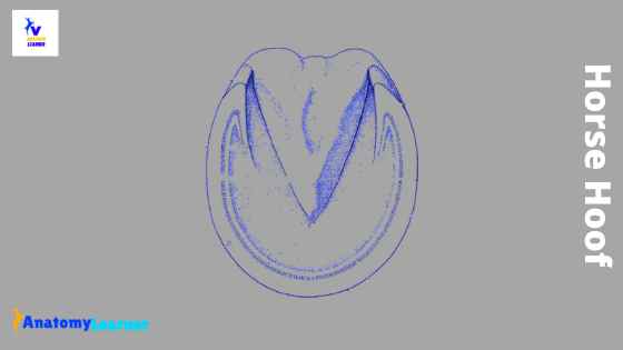
Horse hoof anatomy diagram
Now, I will show you different diagrams of horse hoof anatomy. If you need more updated diagrams on horse hoof, please join anatomy learner on social media.
You may find some mistakes in the labeled diagram of the horse hoof. Please let me known if you found any errors in labeling.
Frequently asked questions on horse hoof.
So, in this part of this article, you will get the answers to the frequently asked question on horse hoof anatomy.
Do horses feel pain in their hooves?
In case of defective hoof trimming or shoeing, horses sometimes may feel pain in their hooves. But proper shoeing is more comfortable for them. You might care about the sensitive laminae of the horse hoof during trimming.
Generally, the wall of the horse hoof does not supply any nerves. So, if you trim the hoof wall, they don’t feel pain in their feet.
What are the parts of the hoof?
Mainly, there are three main parts in a horse hoof: the wall, sole, and frog. Again, the wall of the horse hoof is subdivided into three parts – toe, quarters, and heel. You will also find different structures in the sole and the frog of a horse hoof.
What is the inside of a horse’s hoof called?
As you know, there are three main parts of a horse hoof. In the wall of a horse hoof, you will find a lateral and medial part. This lateral and medial part of the horse’s hoof is known as the quarter.
What is the horn on a horse hoof?
You know the hoof consists of epithelial cells that are more or less wholly keratinized. The keratin of the hoof wall thickens and is cornified to form the horn. The horn makes the outer surface of the horse hoof and protects it from different types of damages.
Does cleaning a horse’s hoof hurt?
It is essential to clean the horse hoof daily. You might know the horse hoof anatomy and the cleaning process of the horse hoof. If you know how to use the simple hoof picker to clean the horse hoof, your horse will not get any pain.
Is a horse’s hoof like a nail?
Yeah, you may consider the horse’s hoof like a nail. The outer part of the horse hoof is keratinized and cornified.
Conclusion
I hope this simple article might help you to get an idea of the horse hoof anatomy. I always request you to use the actual horse hoof sample along with the labeled diagrams.
Again try to identify all of the structures from the actual sample of horse hoof and then go through the details anatomy. If you need any other information related to horse hoof, please let me know. I will update the horse hoof labeled diagram in the future.