Dog Abdomen Anatomy – Abdominal Muscle and Organs
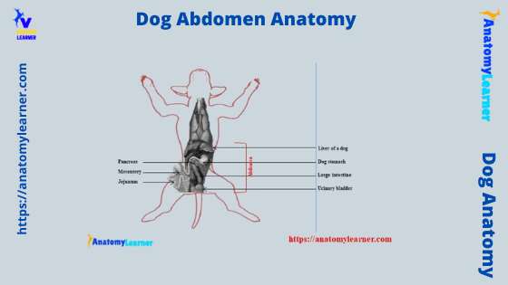
The dog abdomen anatomy consists of boundaries of the abdominal cavity with its organs and associated structures. This article might help you with the details of anatomical facts of the abdomen, both male and female. First, I will try to show you the exact boundary of the dog’s abdomen so that you may also identify … Read more