Structure of a Long Bone – Shaft with a Labeled Diagram
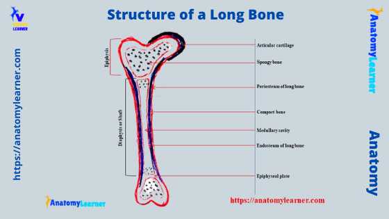
Before studying the animal appendicular skeleton, you might have good knowledge about the parts and structure of a long bone. You will find three major parts (body and extremities) and almost seven major structures in a long bone of an animal. This article will provide detailed information on a long bone’s gross and microscopic features … Read more