
In pituitary gland histology of animal, you will find the adenohypophysis and neurohypophysis parts. You might identify the different cells and structures from the adenophpophysis and neurohypophysis of pituitary gland slide under light microscope.
Hi, do you want to identify the pituitary gland histology slide under light microscope? In this article I am going to show you the important structures from pituitary gland histology with real slide. I will discuss on the different parts of adenohypophysis and neurohypophysis along with their identifying characteristics.
It is so important to know the anatomy of pituitary gland from different animal. At the end of this article I will share few pituitary gland slide labeled diagram with you.
If you want to know about the histological features of pituitary gland with most important identification points then you may continue this article.
Pituitary gland histology
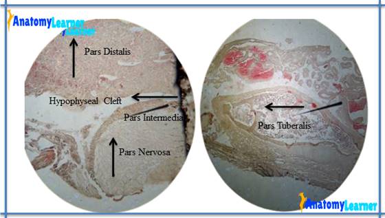
Pituitary gland or hypophysis cerebri is one of the most important endocrine gland in animal body. This gland lies in the body depression of the sphenoid bone (sella turcica) of skull. You will find the two main part of pituitary gland – adenohypophysis and neurohypophysis. Again, you will find pars distalis, pars intermedia, pars tuberalis under adenohypophysis and pars nervosa, infundibulum, median eminence under neurohypophysis part of pituitary gland.
I will discuss the histological features from both adenohypophysis and neurohypophysis of pituitary gland histology slide. But now I would like to provide you a list of structures that you should identify from the pituitary gland.
#1. Capsule of pituitary gland
#2. Pars distalis part of pituitary gland (identification of acidophilic, basophilic and homophobes cells)
#3. Sinusoid in pars distalis
#4. Hypophyseal cleft in pituitary structure
#5. Pars itermedia of pituitary gland
#6. Unmyelinated nerve fibers of pars nervosa
#7. Pituicytes in pars nervosa of pituitary gland
Fine, let’s find these structures from the pituitary gland histology labeled slide diagram.
Pituitary gland histology identification points
So, do you want to identify pituitary gland slide under light microscope? I am going to share the important identification points of pituitary gland histology slide.
#1. Pars distalis consists of groups or cords of cells (homophobes and chromophils)
#2. Numerous sinusoidal capillaries are present in between the groups of cells
#3. Pars intermedia contains colloid filed follicles and it is separated from pars distalis by hypophyseal cleft
#4. Presence of unmyelinated nerve fiber and pituicytes in pars nervosa of adenohypophysis part of pituitary gland
Anterior pituitary gland histology or adenohypophysis
You already know, adenophypophysis has three different part – pars distalis, pars intermedia and pars tuberalis. Now I am going to describe every single parts of adenohypophysis histology from pituitary gland.
In adenohypophysis or anterior pituitary histology, you will find the cluster or group of cells with sinusoidal capillaries. Based on routine stained of stored secretion the cells of pars distalis has classified as chromophlis and chromophobes.
Again the chromophils of pars distalis are two different types – acidophilic and basophilic in nature. These chromophilis cells varies with, size, number, shaped and depending on gender, animals, age and sex. The cytoplasm of these chromophlis of adenohypophysis stained with reddish color.
In acidophlic cells you will find the somatotrophs and lactrotrophs (in orange G dye stain). The secretory granules of lactotrophs is larger than somatotrophs cells granules and they are lightly stained.
In basophilic cells of pars distalis you will find the thyrotrophs and gonadotrophs. The gonadotrophs cells of pars distalis are relatively small and stained with aldehyde thionine dye.
Chromophobes are the small and poorly stained cells with dye used to identify the basophilic and acidophilic cells of pars distalis.
The structure of pars intermedia is different in different animals. In some animal, parenchyma of pars intermedia arranged as simple columnar epithelium, you may also find colloid filled cysts or follicle in some animals. The pars tuberalis of adenohypophysis consists of cell clusters and formed a folded tissue with small cysts (occasionally).
Neurohypophysis histology of pituitary
You know neurohypophysis consists of another different three parts and this neurophypohysis does not contain any secretory cells. In this part of pituitary gland histology, you will find unmyelinated nerve fibers and highly branched glial cells known as pituicytes cell. These glial cells are the modified astrocytes of neural lobe.
Hormone release from the axon terminal in the neural lobe that are synthesized in neural perikarya of magnocellular hypothalamic supraoptic and paraventricular nuclei
If you want to learn the anatomy of pituitary gland with their functions of different parts then find the article at veterinary gross anatomy learning section. Hope you will learn the basic anatomy of pituitary gland of animal with labeled pictures
Hypophysis cerebri histology slide picture drawing and labeling
Fine, now I am going to share the pituitary histology slide drawing picture with you. Here in this picture you will find all the structures that I have enlisted before. If you found nay mistake in pituitary slide picture labeling then let me inform.
Again, if you want to get more slide pictures of pituitary histology then get them at here in anatomy learner social media.
If you want then you might learn the other histological features (identifying characteristics) of different endocrine gland and lymphoid organs or other different organs from animal body from anatomy learner.
#1. Histological features of pancreas under light microscope with labeled slide pictures
#2. Identifying histological characteristics of thymus of animals under light microscope
#3. Identify the different histological features or structures from animal ovary histology slide
#4. Histology of adrenal gland cortex and medulla with real histology slide and pictures
Conclusion
Hope this guide was helpful to learn pituitary gland histology. Now you will able to identify the anterior and posterior pituitary gland histology slide under microscope with important identification points.
Don’t forget to join social media of anatomy learner for more update pituitary gland histology labeled pictures and diagram.
Share this article with your friends who want to learn the histology of pituitary with labeled diagram and real pictures.
If you find any mistake on the labeled pituitary slide picture please let me inform. Many thanks for staying with anatomy learner and learn gross anatomy and histology with me.