Where is Stratified Cuboidal Epithelium Found

Where is Stratified Cuboidal Epithelium Found

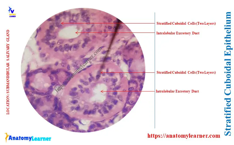
The stratified cuboidal epithelium has a limited distribution in the animal body. This guide will help answer the question: Where is stratified cuboidal epithelium found? Quick answer: the stratified cuboidal epithelium is found in the excretory ducts of salivary glands, sweat glands, and developing ovarian follicles. This type of cuboidal epithelium is also found in … Read more

Skeletal Muscle Under Microscope with Labeled Diagram

Skeletal Muscle Under Microscope

The skeletal muscle under a microscope shows long, cylindrical, unbranched fibers and flat nuclei. Each skeletal muscle fiber comprises compactly packed long myofibrils that arrange parallel to the long axis. This article might help you to understand the microscopic features of the skeletal muscle with the labeled diagram. Again, you will find the complete list … Read more

Stratified Squamous Epithelium Under Microscope with Labeled Diagram

Keratinized Stratified Squamous Epithelium Under a Microscope

The stratified squamous epithelium under a microscope shows the multiple layers of cells where the superficial one is flattened. You will also find the basal columnar cells in this stratified squamous epithelium. Quick summary of stratified squamous epithelium: in this type of epithelium, you will see multiple layers of cells where the basal layer is … Read more

Simple Columnar Epithelium Under a Microscope with Labeled Diagram

Simple Columnar Epithelium Under a Microscope

The simple columnar epithelium under the microscope shows a single layer of cells resting on the basement membrane. Here, I will show you the microscopic features of the simple columnar epithelium with their location and labeled diagram. Quick summary of simple columnar epithelium: these are the single layer of cells with a greater height than … Read more

Cardiac Muscle Under Microscope with Labeled Diagram

Cardiac Muscle Under a Microscope Labeled 400X Diagram

The cardiac muscle under a microscope shows a short cylindrical fiber with a centrally placed oval nucleus. You will find some unique features in cardiac muscle that will help you to differentiate it from skeletal and smooth muscles. Quick answer: the cardiac muscle microscope slide shows the cylindrical fiber with 1 or 2 nuclei. The … Read more