Lymphocytes Under Microscope with Labeled Diagram

Lymphocytes Under a Microscope with Labeled

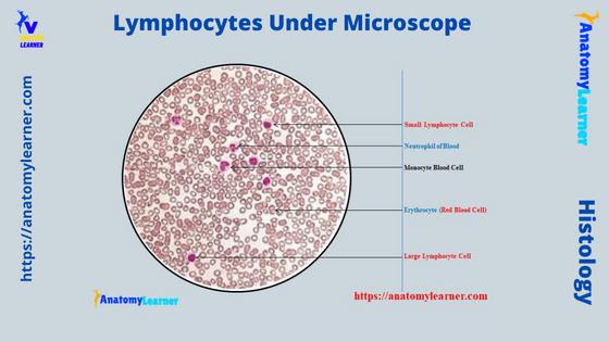
Lymphocytes under a microscope show a round to slightly indented nucleus with clumped heterochromatin. You know these lymphocytes are agranulocytes and form the second-largest population of white blood cells. This article will show you the structure (morphology) of different types of lymphocytes (small, large, B, and T) under light and electron microscopes. So that after … Read more

Adipose Tissue Under Microscope with Labeled Diagram

Adipose Tissue Under Microscope Labeled

The adipose tissue under a microscope shows an aggregation of fat cells or adipocytes. You will find this adipose tissue subcutaneously throughout the body except over the eyelid, auricle, and other parts of the animal’s body. The adipose tissue is considered the specialized connective tissue which plays an essential role in energy homeostasis. Microscopically, you … Read more

Prophase Under Microscope – from Mitosis and Meiosis Stages

Prophase Under Microscope Labeled Diagram

The prophase under a microscope shows the gradually becoming condensed chromatin, resulting in the formation of the individual chromosome. You know this prophase is the first stage of mitosis cell division which may quickly identify with the help of a light microscope. Again, you will also see the prophase 1 and prophase 2 stages in … Read more

Tongue Under Microscope with Labeled Diagram

Tongue Under Microscope Labeled

The tongue under a microscope shows a core of crisscrossing skeletal muscle bundles and a peripheral mucous membrane. A stratified squamous epithelium covers the mucous membrane of the tongue and contains 4 types of papillae. Again, each tongue papilla possesses a connective tissue core that covers the epithelium. Some of these papillae of the animal’s … Read more

Binocular Microscope Anatomy – Parts and Functions with a Labeled Diagram

Binocular microscope anatomy labeled diagram

It is essential to know the binocular microscope anatomy for the first-year veterinary student before starting histology learning. This article might help you know different parts of the binocular compound microscope with a labeled diagram. Most first-year students don’t know how perfectly set up a slide under the light microscope. So, I planned to tell … Read more