Colon Histology Slide with Labeled Diagram

Colon histology slide labeled diagram

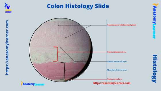
The colon histology slide possesses the typical four layers of a tubular organ – mucosa, submucosa, muscularis, and serosa. But, there are no permanent plica circularis and villi in the colon slide as found in the different segments of the small intestine. You will also see a thicker tunica mucosa in the colon slide because … Read more

Hair Under a Microscope – Features of Hairs Shaft and Follicles with Labeled Diagram

Hair shaft under a microscope labeled

Hair under a microscope shows a hair follicle and a cylindrical hair shaft. In a hair shaft, you will find columns of keratinized cells organized into three layers – medulla, cortex, and cuticle. Again, the hair follicle is the epidermis structure that develops as down growth of the epidermis into the dermis and possesses different parts.  … Read more

Skin Under Microscope

Skin under microscope labeled

The skin under a light microscope shows two distinct layers – epidermis and dermis. In the case of thin skin, the epidermis is very thin and lines with the keratinized stratified squamous epithelium. You will also find five different cells layers in the epidermis of both thick and thin skin under a light microscope.  The dermis of thick and … Read more

Pseudostratified Columnar Epithelium under a Microscope with a Labeled Diagram

Pseudostratified columnar epithelium

The pseudostratified columnar epithelium comprises a single layer of cells but seems to be multilayered. It is because different cellular heights and nuclei are also placed at a different levels. I will show you the pseudostratified columnar epithelium under a light microscope with its identifying points and labeled diagram.  I will also tell you the location of pseudostratified columnar … Read more

Simple Squamous Epithelium under a Microscope with a Labeled Diagram

Simple squamous epithelium under microscope labeled

Simple squamous epithelium under a microscope consists of a single layer of thin, flat, and scale-like cells. These cells are joined together by an intercellular junction and rest on the basement membrane, whose thickness depends on the location. Here, I will show you what simple squamous looks like under a light microscope.  Again, I will also tell you almost … Read more

Neuron under Microscope with Labeled Diagram

Neuron under microscope labeled

The structural and functional unit of the nervous system is the neuron that may easily observe under a light microscope. Neurons may vary considerably in size, shape, and other features. Here, I will provide details information and identifying points so that you may easily identify the neuron under a microscope.  A neuron consists of a cell body that gives … Read more