Ruminant Reticulum Histology Slide with Labeled Diagram

Ruminant reticulum histology slide

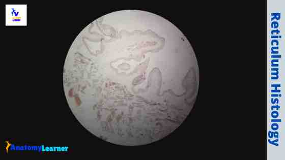
The reticulum is a non-glandular part of the ruminant stomach. In the ruminant reticulum histology, you will find some permanent interconnecting mucosal folds known as reticular crests. I will show you every essential histological feature from the ruminant reticulum histology slide with labeled images.  I will also show you some differences among the three different parts of the ruminant … Read more

Cecum Histology Slide with Labeled Image and Diagram

Cecum histology slide labeled

The cecum histology slide follows the general pattern of the small intestine with some exceptional features. You may find a variation in the size of the gross cecum among the different species. But, in all domestic mammals, the cecum histology slide shows a substantial number of lymphatic nodules scattered throughout its length. Here, I will show you all the … Read more

Parathyroid Gland Histology with Microscope Slide Image and Labeled Diagram

Parathyroid gland histology slide

There are two or more pairs of parathyroid glands located on the posterior surface of the thyroid gland. You will find two main types of cells (chief and oxyphils) in the parathyroid gland histology slide. Here in this article, I will show you all the structures of a parathyroid gland with a microscope slide image and a … Read more

Tonsil Histology Slide with Labeled Diagram – Histological Features of Palatine and Lingual tonsils

Tonsil histology slide microscope image

Near the junction of the oral cavity and the pharynx, you will find several collections of lymphoid tissue that refer to the tonsil. In tonsil histology, different essential structures like tonsillar crypts, lining epithelium, numerous lymphatic nodules, and dense connective tissue capsules should be identified under a light microscope.  I will show you all these structures from … Read more

Blood Histology Slides with Description and Labeled Diagram

Blood histology slides

The blood is a specialized connective tissue that is fluid and circulates through the vascular channel. In the blood histology slide, you will find different types of cells with their specific features. This might be a short article where I will show you all the cells from the blood microscope slide with a labeled diagram and actual … Read more

Urinary Bladder Histology with Microscopic Slide Image and Labeled Diagram

Urinary bladder histology slide images

The urinary bladder is a muscular sac-like structure that temporarily stores urine and discharges out prediocally through the urethra. In the urinary bladder histology, you will find three or four defined coats like the tubular organs. Here, I will show you all the layers from the urinary bladder histology slide with microscopic images.  You will also get information on … Read more